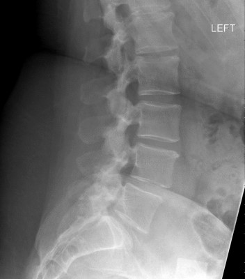
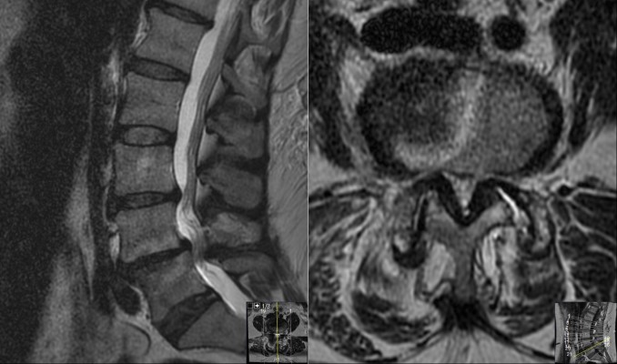
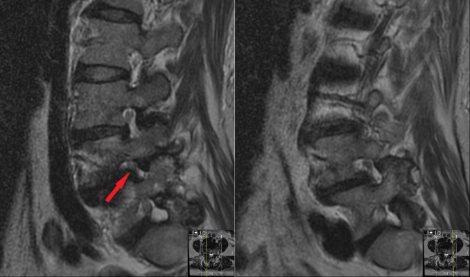
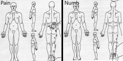
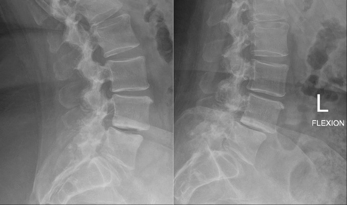
This is a 46 year-old female with back and right leg pain. She reported back pain for 4 years and leg pain severe for about 1 year. She had aching in her back and burning in the right, more than left, calf with walking. She also had numbness and tingling into the right calf and foot and tripping due to dragging her right foot. On exam, she has loss of sensation in the right foot and some weakness of the right ankle.
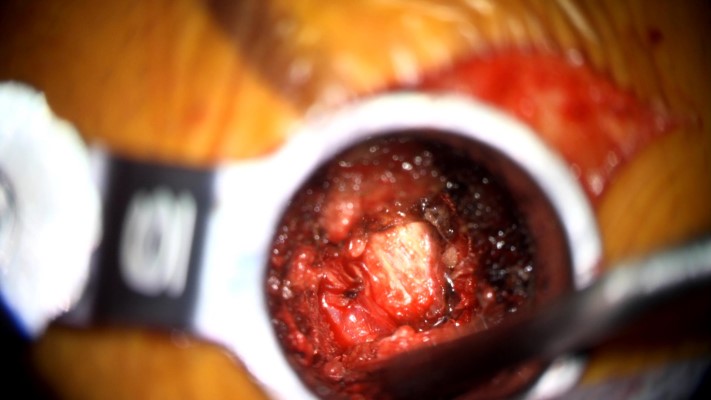
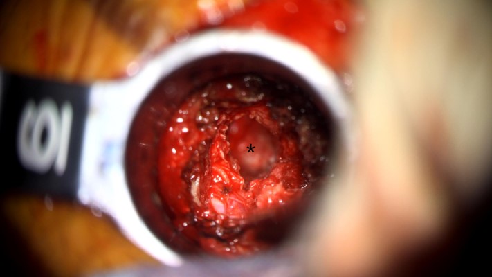
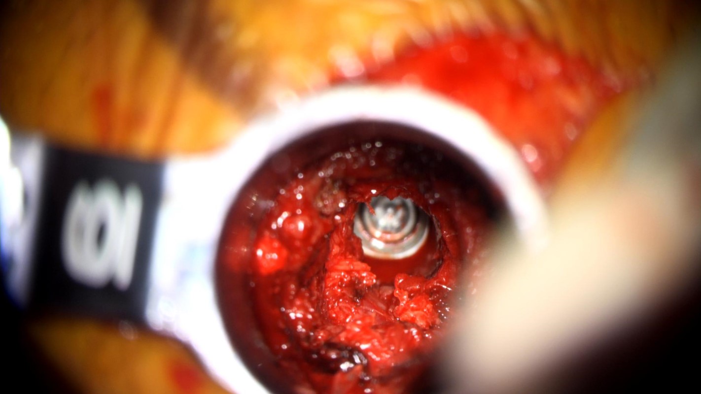
Intraoperative video captured through the operative microscope. It shows the use of a high-speed drill in the tubular retractor removing bone to decompress the nerve in the neural foramen, and gain access to the disc space.
- All
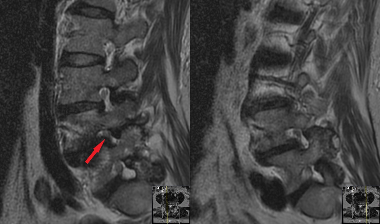
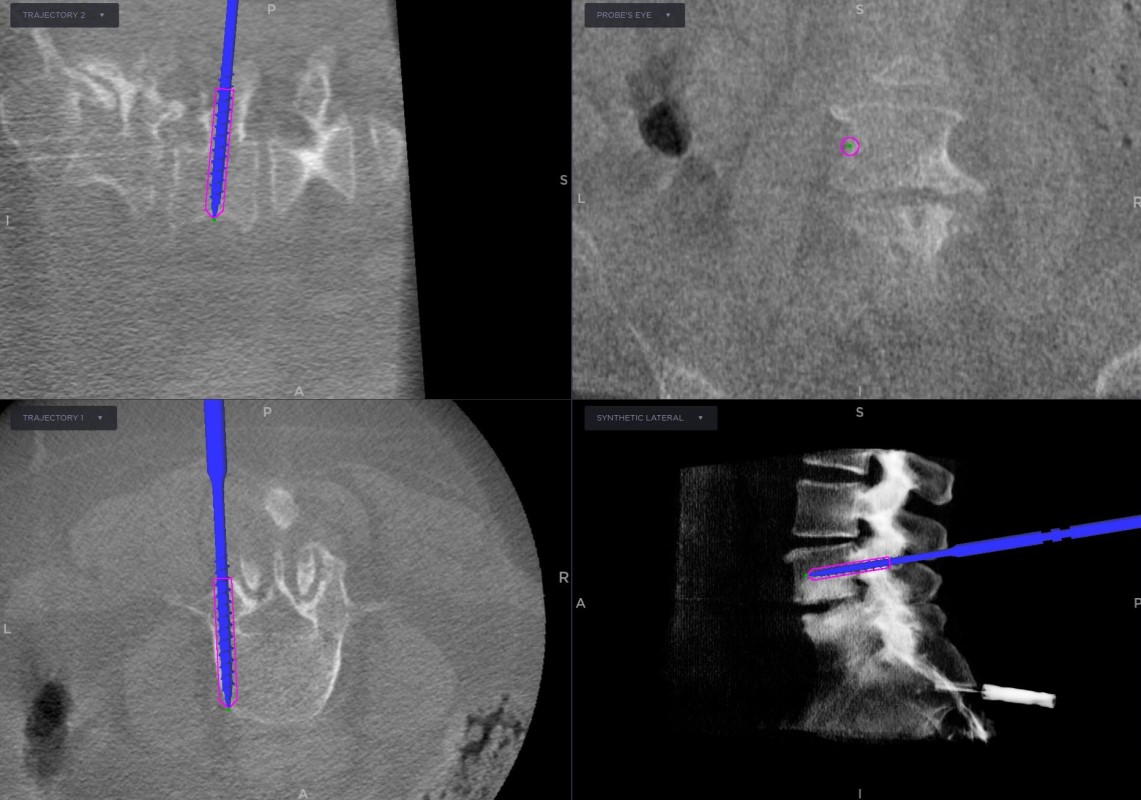
- Pre-Op
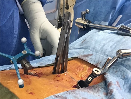
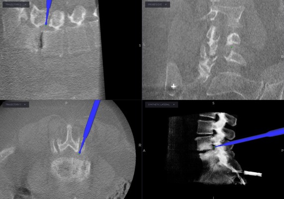
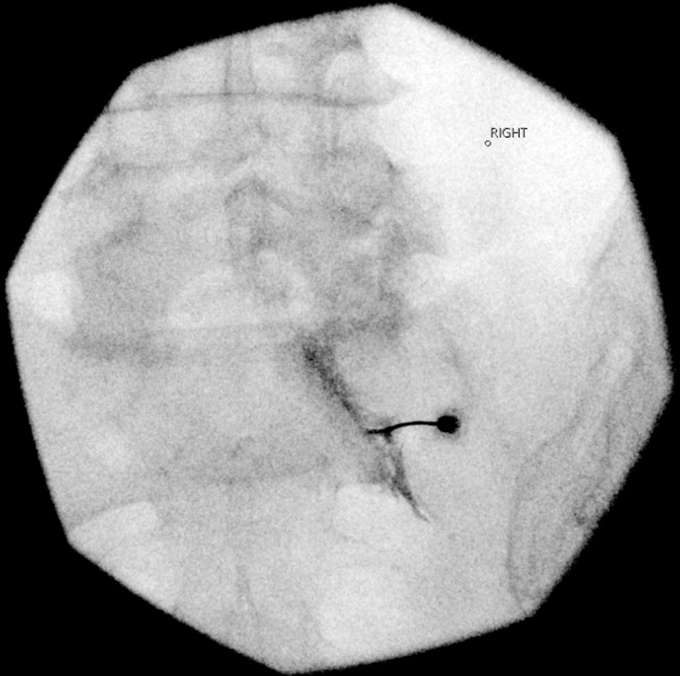
- Intra-op
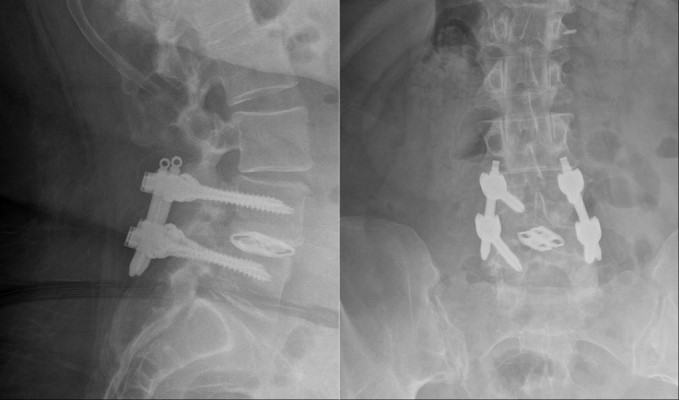
- Post-op


