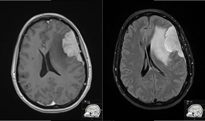
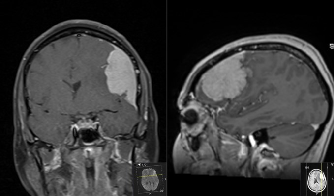
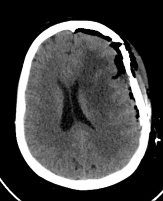
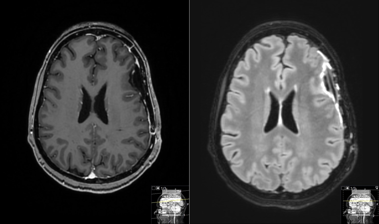
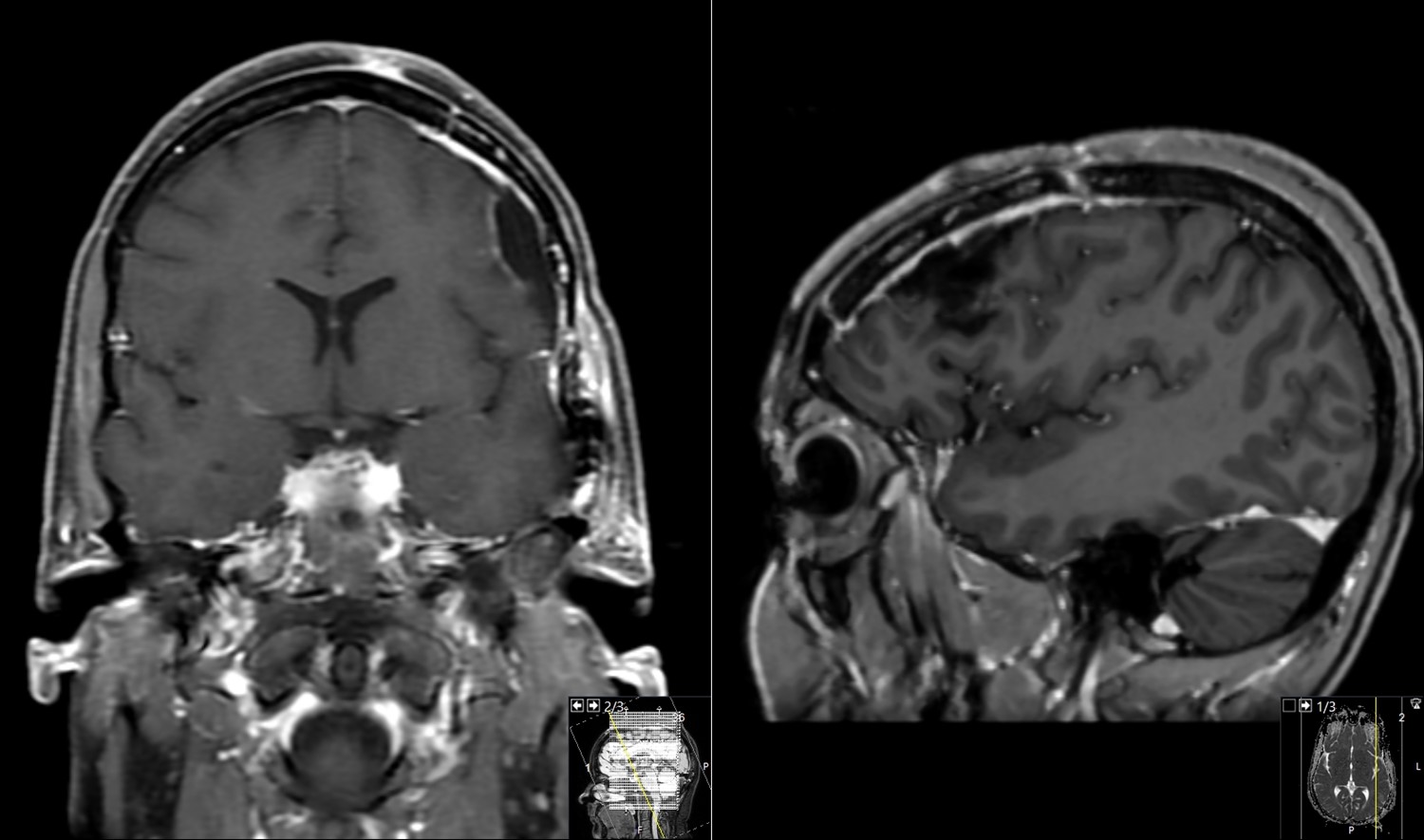
This a case from a 52 year old woman presenting to ER with new onset seizure.
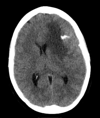
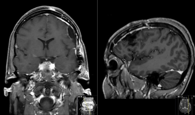
CT scan of the head showing an abnormality in the left frontal region (top right of image). It includes a bright white area consistent with calcification. The adjacent brain is darker on that side due to edema, and the midline structures of the brain are pushed over top the right (called midline shift).
- All
- Pre-Op
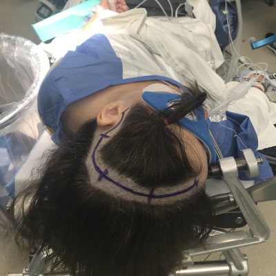
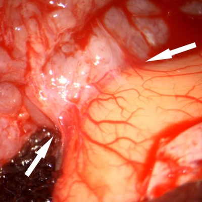
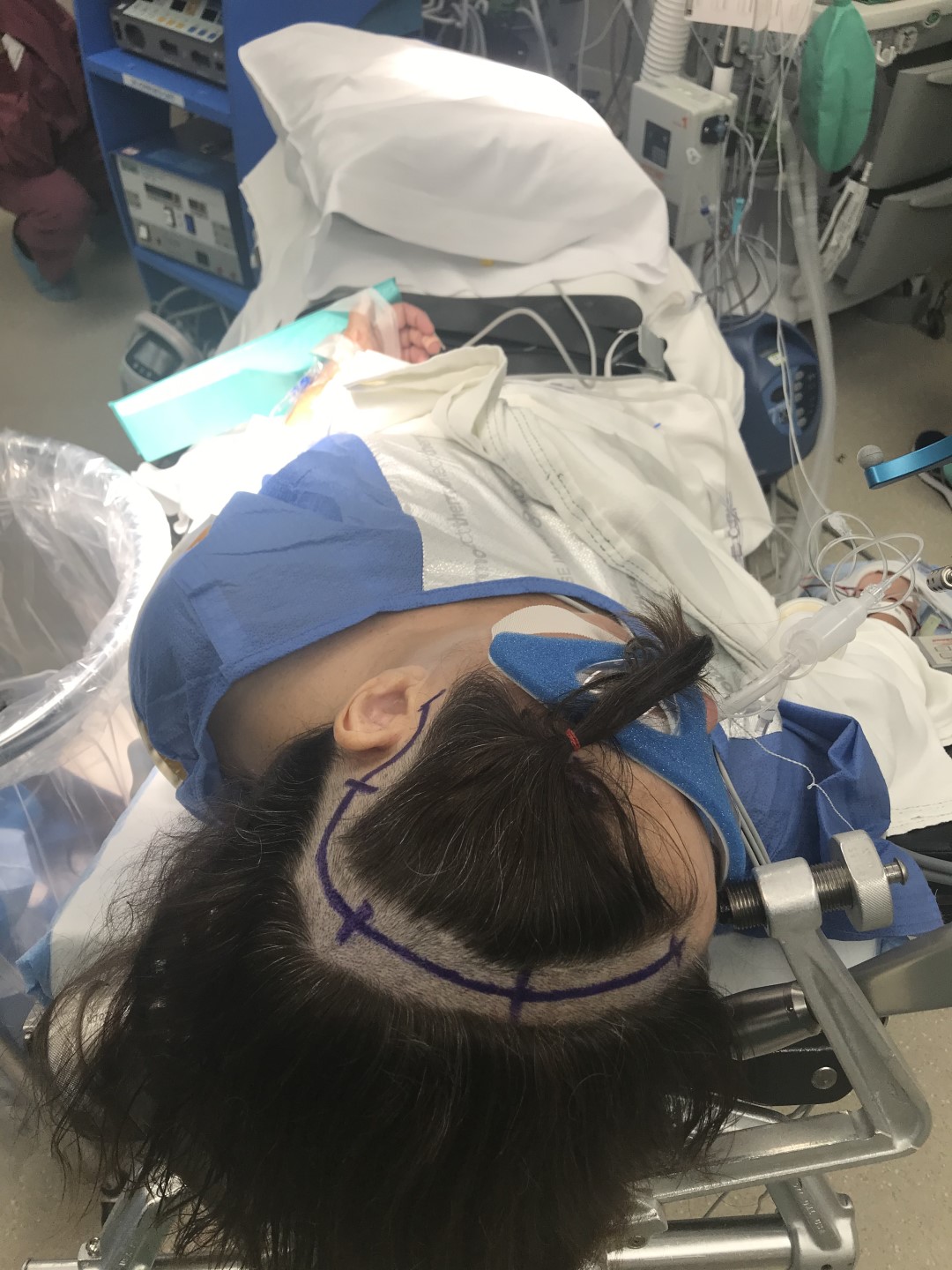
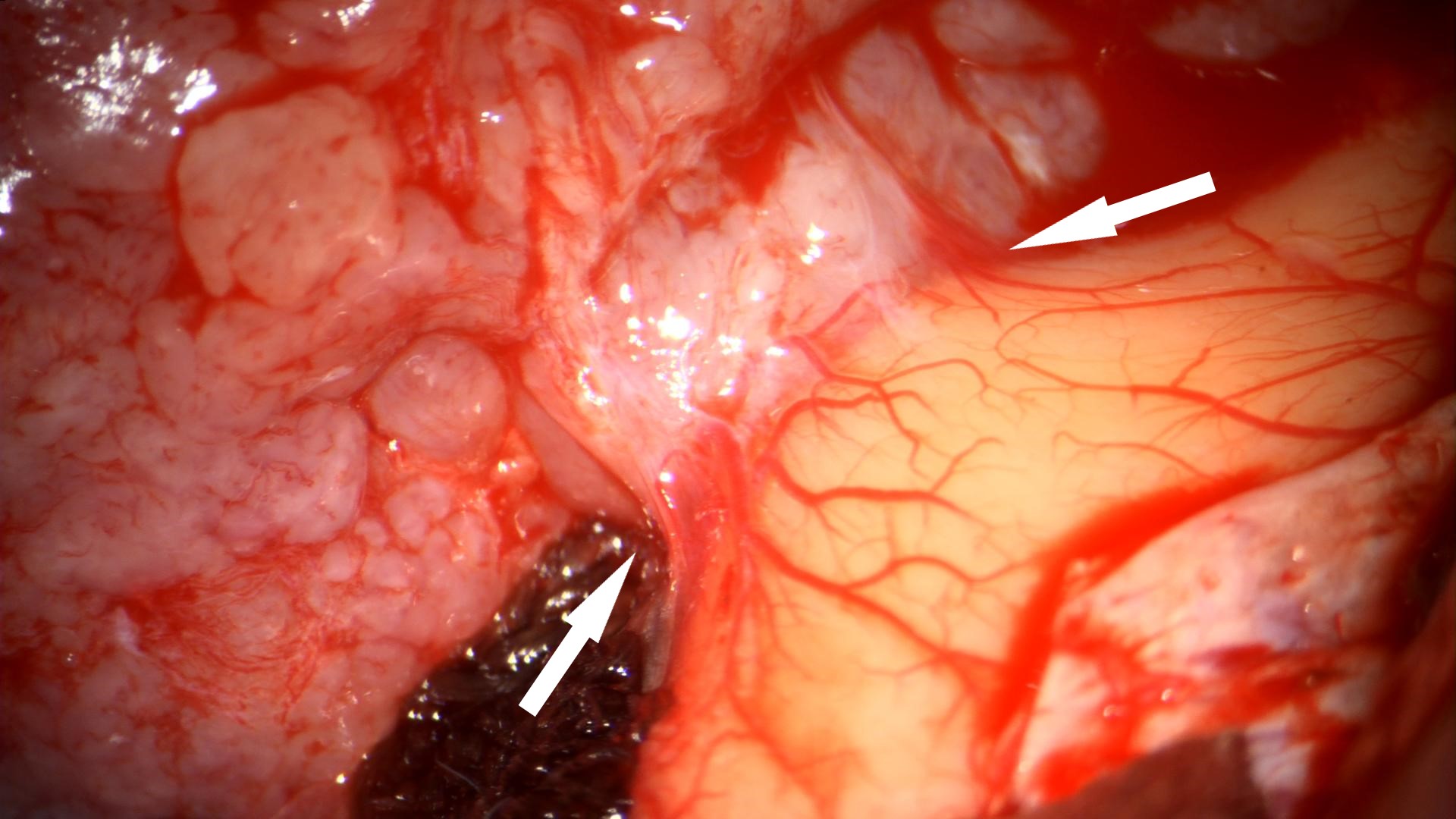
- Intra-op
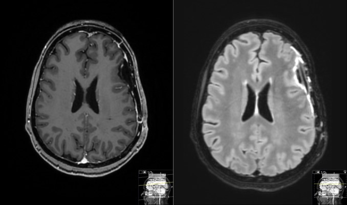
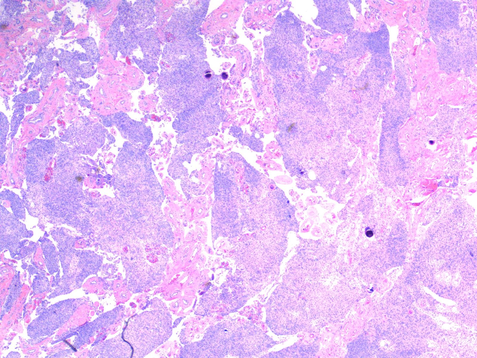
- Post-op