This is a 57 year-old who presented with a primary complaint of pain in his left gluteal region (buttocks) that radiates down to the back of his left thigh. He had no numbness, tingling, or weakness. He did not respond to an epidural steroid injection. His neuro exam was unremarkable (no sensory or motor deficit). The Musculoskeletal exam with signs of left SI joint mediated pain. The patient engaged in physical therapy for back and hips and returned to interventional pain management physician who performs left SIJ injection which improved his gluteal pain from 8/10 to 5/10.

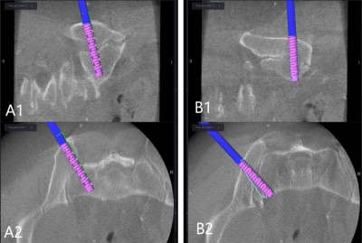
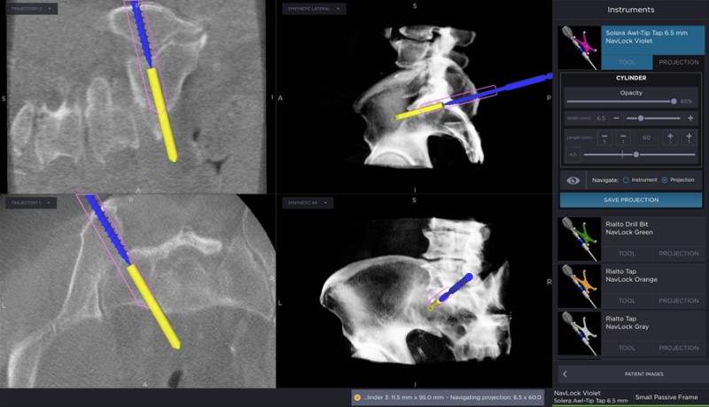
Image captured from intraoperative image-guided navigation workstation showing the trajectory used to place the first screw across the left SI joint. Note the planned trajectory of the screw (pink outline), the position of the tap (blue) and the trajectory upon which the tap is being advanced (yellow).
- All
- Pre-Op
- Intra-op
- Post-op
