This a case of a 66 year old female who presented with leg greater than back pain for more than one year. The pain radiated down the back of the legs with numbness and tingling in her calves. Her symptoms were worse with walking and standing and better with rest. Treatment included injections and physical therapy.
- All
- Pre-Op
- Intra-op
- Post-op
 CT abdomen from 3 years prior (2017). It shows normal alignment between L4 and L5.
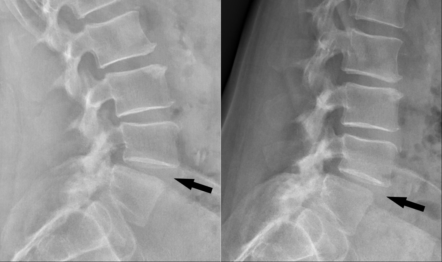
 Preop dynamic x-rays. These side-view images show a malalignment between L4 and L5 (called a spondylolisthesis. There is a slight change in the relationship between L4 and L5 from bending back (left, aka extension) and bending forward (right, aka flexion). Arrow points to L4/5 disc space.
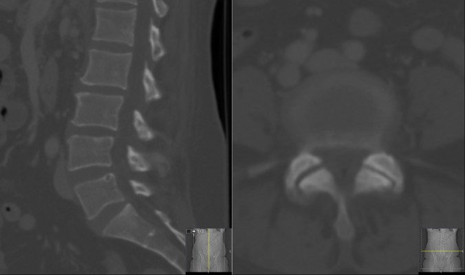
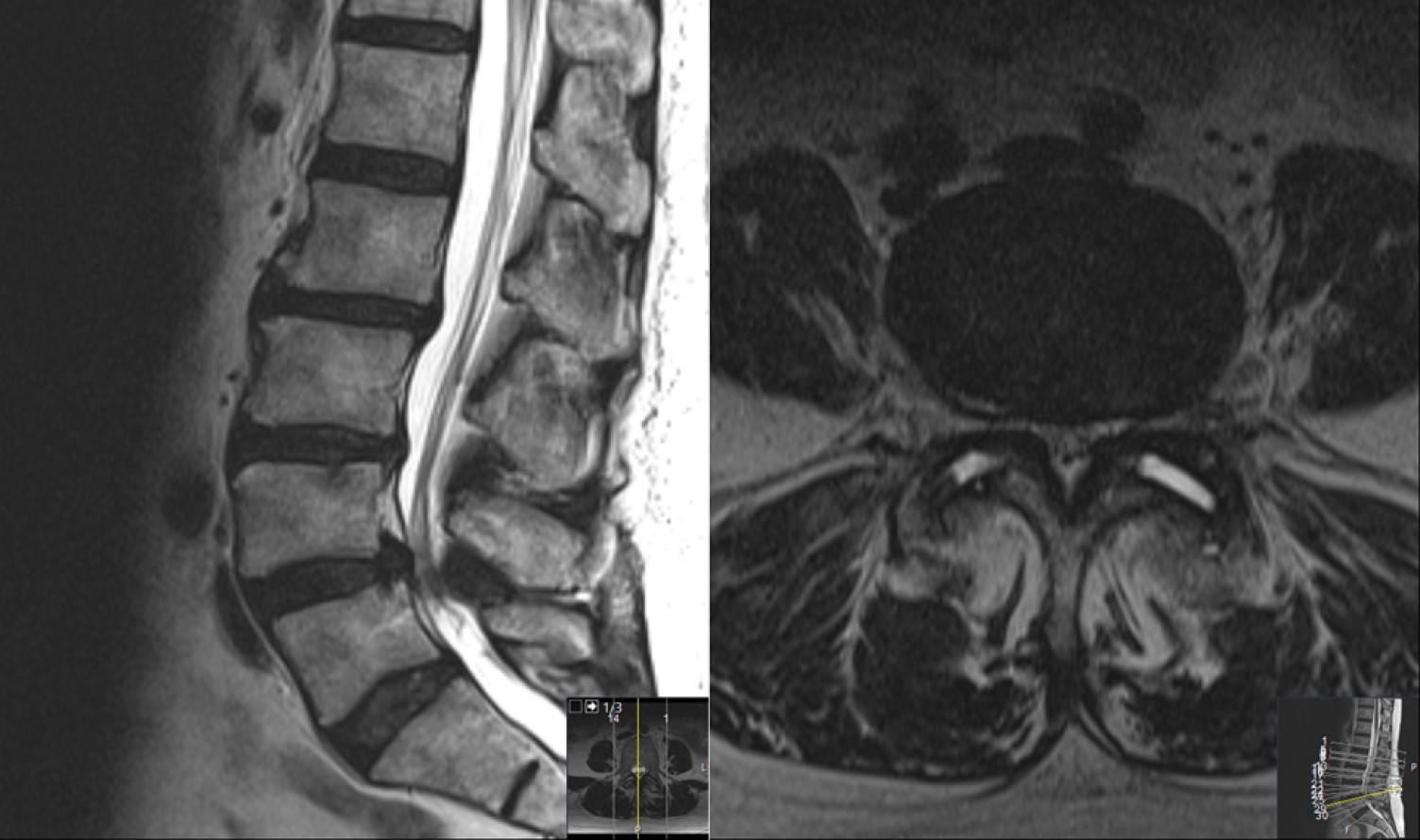
 Preop MRI: the side view image (left, aka sagittal) shows the L4/5 malalignment and pinching of the nerves, while the cross-cut image (right, aka axial) shows severe arthritis and the nerves being compressed (aka stenosis)
 : Intraoperative image acquired through the surgical microscope- angling in from the left side. The surgical approach has removed soft tissue overlying the spine. The image shows bone on the left side covering the underlying nerves
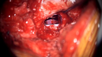
 bone has been removed, and the covering of the underlying nerves (the dura) has been exposed
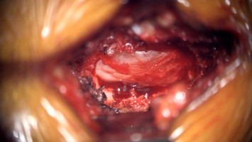
 the dura is being retracted exposing the intervertebral disc which lies underneath (with some veins attached to its surface)
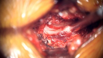
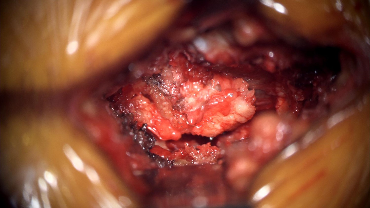
 the dura is again being retracted, the disc has been removed, and a metal implant (magenta in color) can be seen deep in the disc space.
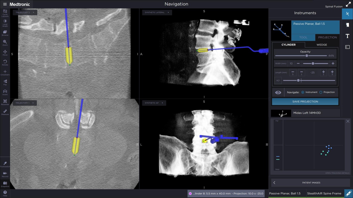
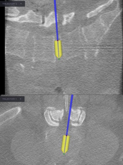 Image Capture taken from the Stealth navigation workstation during surgery. It shows the image-guided probe within the L4/5 disc space being used to correctly size the interbody spacer and guide it into place.
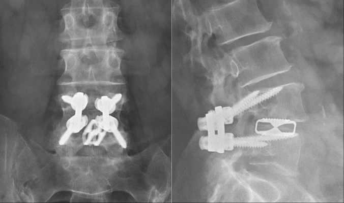 PostOp x-rays: taken 6 months after surgery, these front-on (left, aka AP) and side-view (right, aka lateral) images show the four screw, two rods and cage in position. Compare these to the preop xray (figure2) and note the improved alignment between L4 and L5.
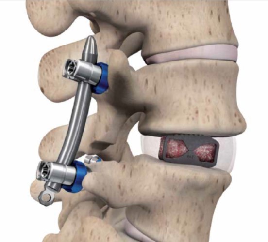
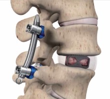 Diagram demonstrating the placement of the spinal hardware as was used in this case (provided by Spine Wave, Shelton, CT)