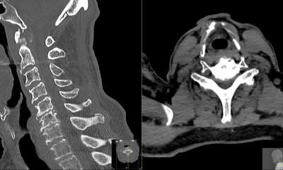
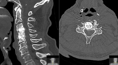
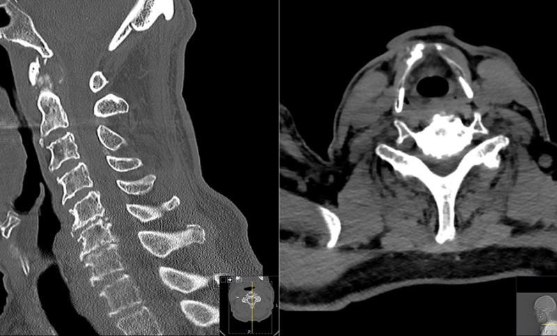
This is an 85 year old male who presented with shock-like sensations shooting into his arms and hands with increasing numbness in hands. He had one month of hand and arm weakness, getting worse rapidly. He was off balance when walking. He is unable to use his hands for fine motor tasks like shaving, buttoning clothes etc.
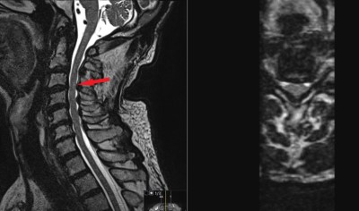
The Musculoskeletal exam was notable for limited motion of cervical spine. Neurological examination with weakness and loss of sensation in hands. He also has abnormal and hyperactive reflexes.
The Musculoskeletal exam was notable for limited motion of cervical spine. Neurological examination with weakness and loss of sensation in hands. He also has abnormal and hyperactive reflexes.
- All
- Pre-Op
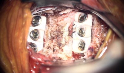
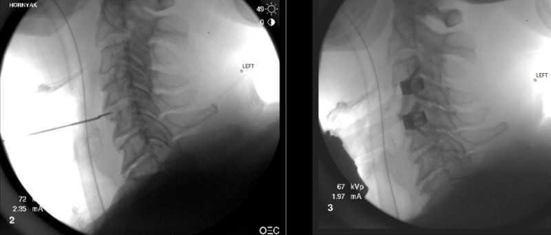
- Intra-op
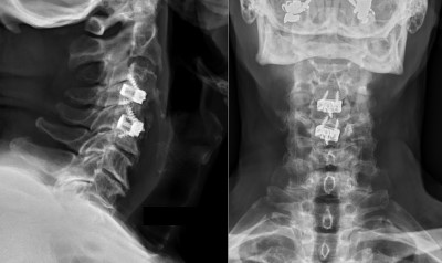
- Post-op

