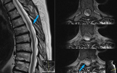
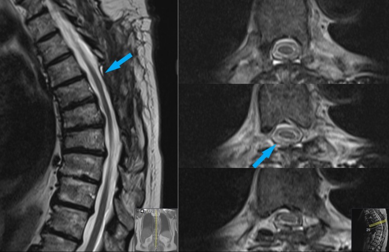
This is a 65 year old female who presents to office with complaint of mid-back pain, progressive over several weeks. She has numbness in her toes for about 2 weeks. She also complains of difficulty walking; feels off balance, but not dizzy. On exam she has mild increased deep tendon reflexes.
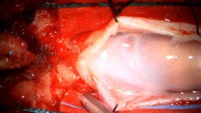
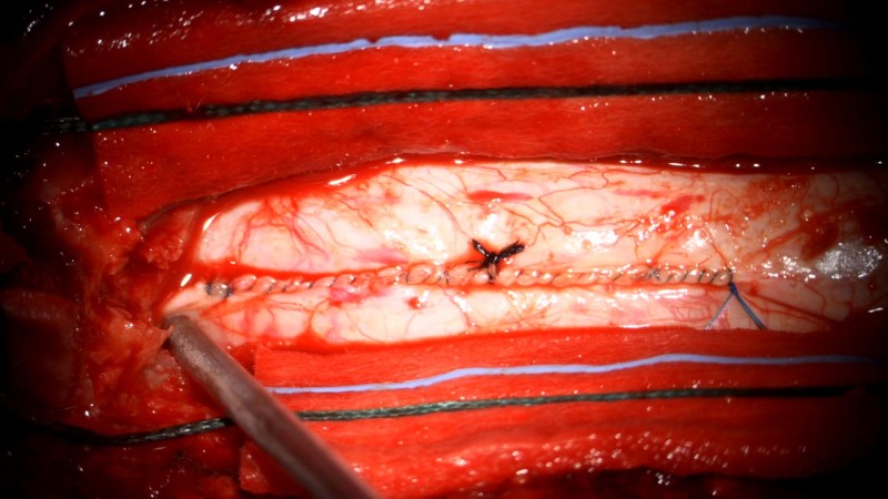
An intraoperative photo taken with the operating microscope. The dura has been opened in the midline and tenting sutures (black string) is holding it open. The thickened arachnoid of the cyst can be seen as the pearly white membrane in the center of the frame (it is normally clear, as is seen on the far right of the frame).
Intraoperative video showing further resection of the cyst. Thick arachnoid bands are divided with microscissors exposing the spinal cord beneath.
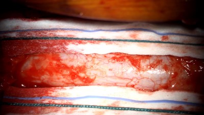
An intraoperative photo showing a single arachnoid band after resection of the arachnoid cyst. The spinal cord is clearly seen beneath.
- All
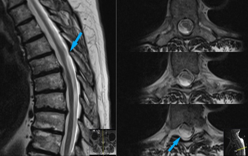
- Pre-Op
- Intra-op
- Post-op

