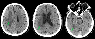
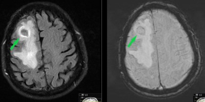
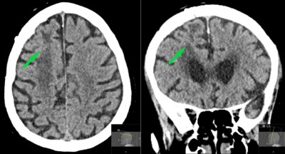
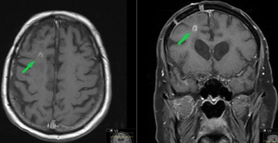
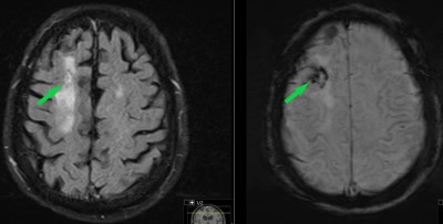
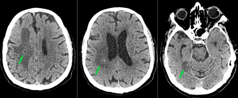
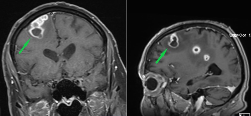
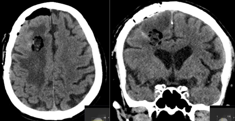
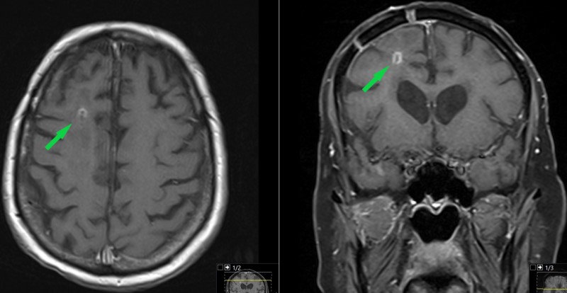
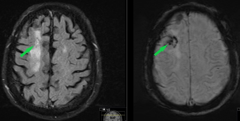
This is a 90 year old male who presented to the ER with several days of progressive left-sided weakness in arm more than leg which led to fall at home prompting presentation to ER. The past medical history is relatively unremarkable other than pancytopenia (low blood cell counts).
  • All
  • Pre-Op
  • Intra-op
  • Post-op
Three images obtained from initial CT head showing multiple lesions in the brain (green arrows)
Axial images from MRI brain. Noncontrast T1-weighted (left) and contrast enhanced T1 (right) images show a ring–enhancing lesion in the right frontal lobe (green arrow).
Coronal (left) and sagittal (right) images obtained from contrast-enhanced, T1 weighted MRI of the brain. Shows large ring-enhancing lesion in the right frontal lobe; this is the same lesion identified in figure 2. Also shows two other lesions on the sagittal image.
Axial images obtained from the MRI of the brain. The FLAIR image on the left shows the cerebral edema throughout the left frontal lobe emanating from the left frontal lesion (green arrow. The SWI image on the right shows that there is no significant hemorrhage associated with the lesion.
Axial (left) and coronal (right) images obtained from CT of the chest. There is a peritracheal mediastinal lesion (green arrow). This lesion was biopsied and found to be insignificant.
Patient underwent craniotomy for excisional biopsy of the right frontal lesion. This image was obtained from the intraoperative navigational workstation. It demonstrates the right frontal lesion and shows a surgical plan for accessing the lesion (thin blue line).
Axial (left) and coronal (right) images obtained from CT head performed immediately postoperatively. Note the changes within the right frontal lobe consistent with recent surgery; also note craniotomy on the coronal image.
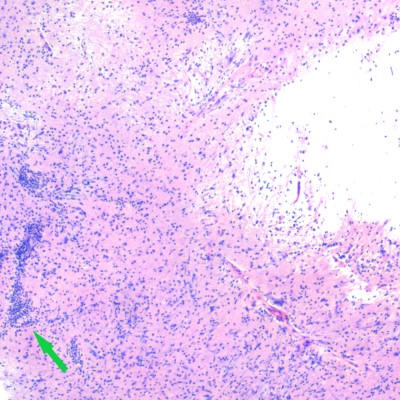
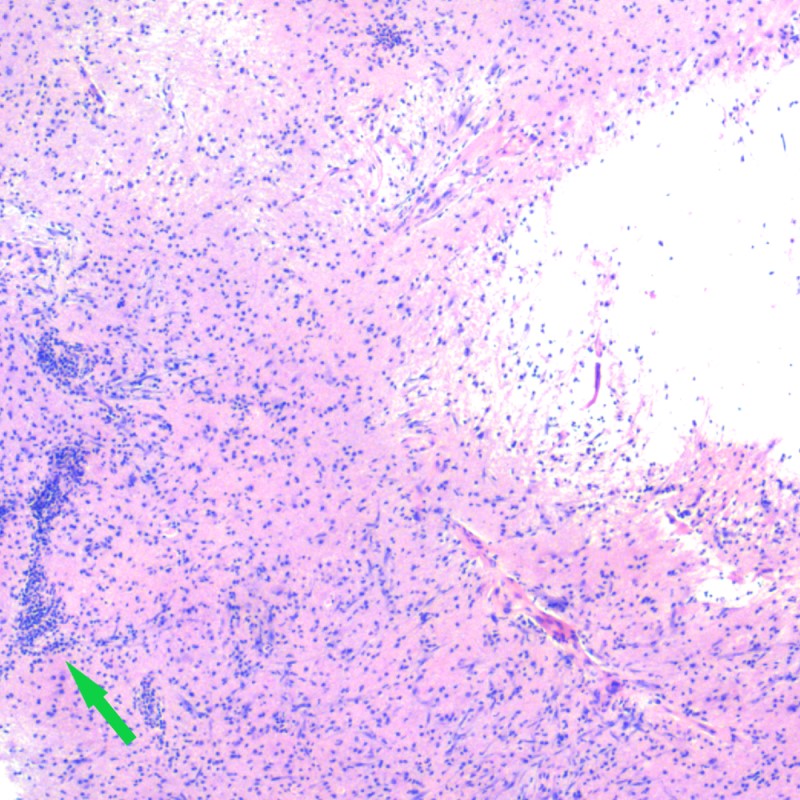
Photo micrograph of patient's pathologic specimen. This H&E stain shows brain tissue (pink) with an inflammatory infiltrate (multiple purple dots throughout the image). There is a cluster of these inflammatory cells above the tip of the green arrow. While abnormal, this does not provide the diagnosis.
Photo micrograph showing FITE acid-fast stain of brain tissue. Again noted are multiple inflammatory cells (blue/purple cells). Also noted are red filamentous forms (one is identified just above the tip of the green arrow) consistent with atypical bacterial infection.
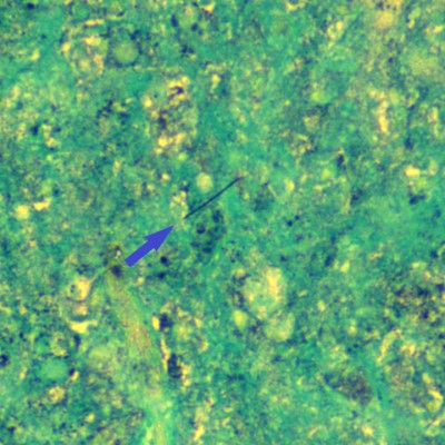
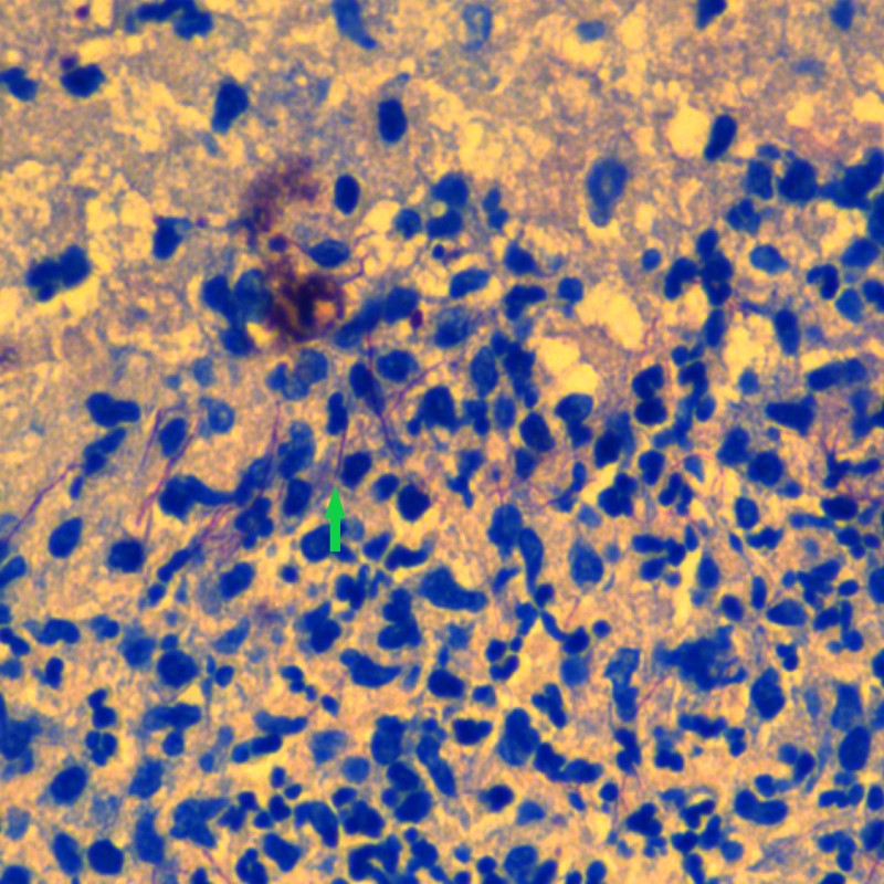
Photo micrograph of GMS (Grocott’s Methenamine Silver) stain of pathologic specimen. It also demonstrates a filamentous microbe consistent with infection (blue arrow). 
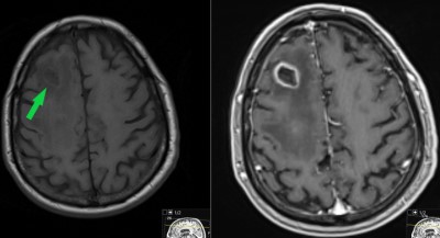
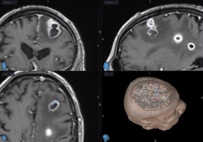
Microbiological specimens collected during craniotomy grow Nocardia araoensis. It is susceptible to multiple antibiotics. Patient is treated with antibiotics.
Axial (left and coronal (right) images obtained from CT head performed approximately 3 months postoperatively. Postoperative changes can still be seen. This is markedly improved compared to the preoperative CT (Figure 1).
Postcontrast T1-weighted images obtained in the axial (left) and coronal (right) planes. These images were obtained approximately 4 months postoperatively. They show near resolution of the right frontal lesion as well as the other lesions within the brain.
Additional images obtained from the MRI brain performed approximately 4 months postoperatively. They also show improvement compared to the preop study with marked improvement in the perilesional edema seen on the FLAIR image (left). The SWI image shows postoperative change.