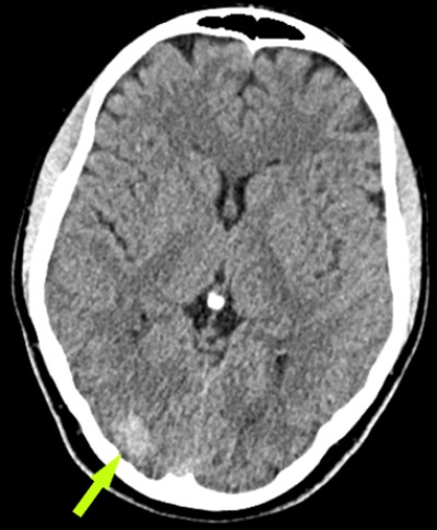
This is a 24 year old female who presented to the ER 2 years prior with first time seizure.
She is admitted to hospital, evaluated, and released.
She did not follow-up with neurosurgery at that time.
One year later, again presents to ER with another seizure; undergoes work-up and is again released.
6 months later she follows up with neurosurgeon.
She has mild, intermittent visual disturbance
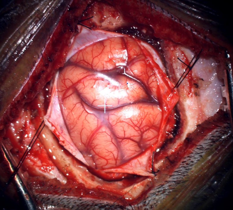
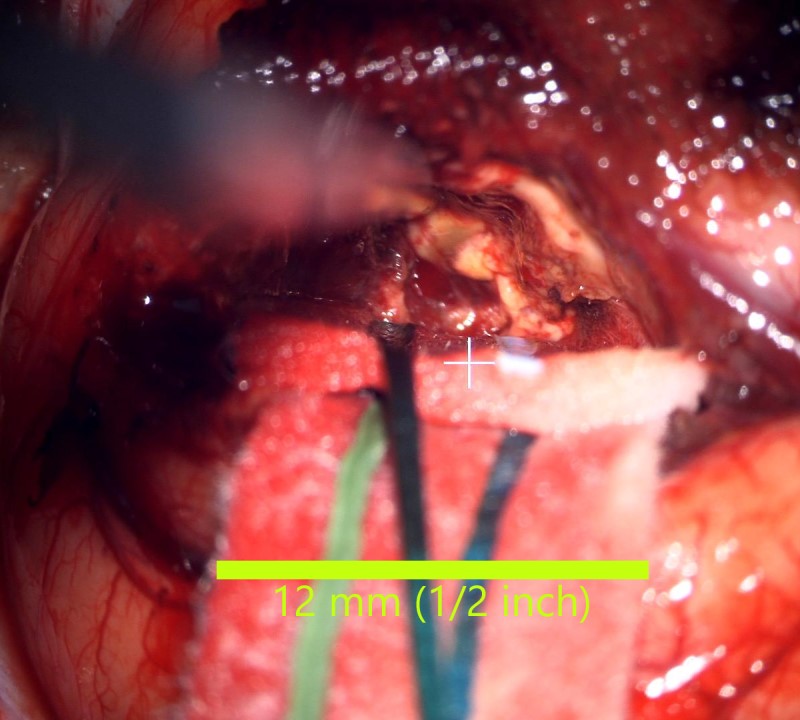
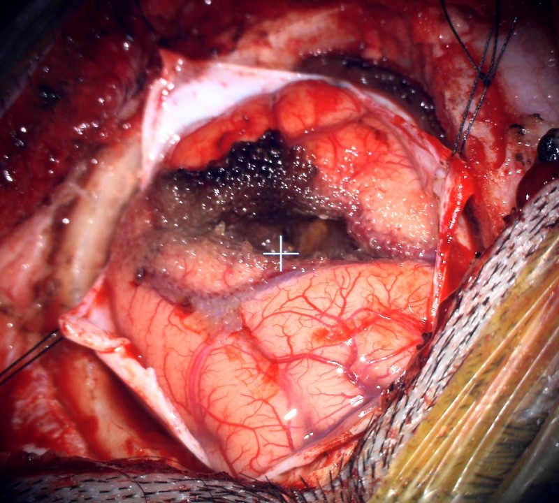
Intraoperative video showing final removal of the lesion.
- All
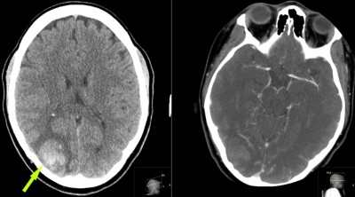
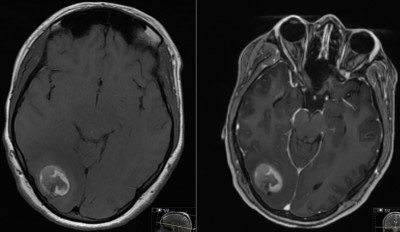
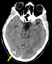
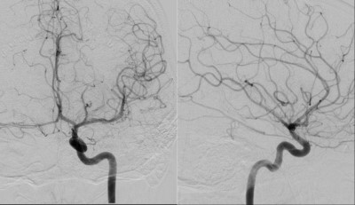
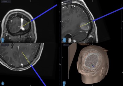
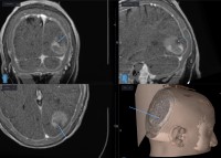
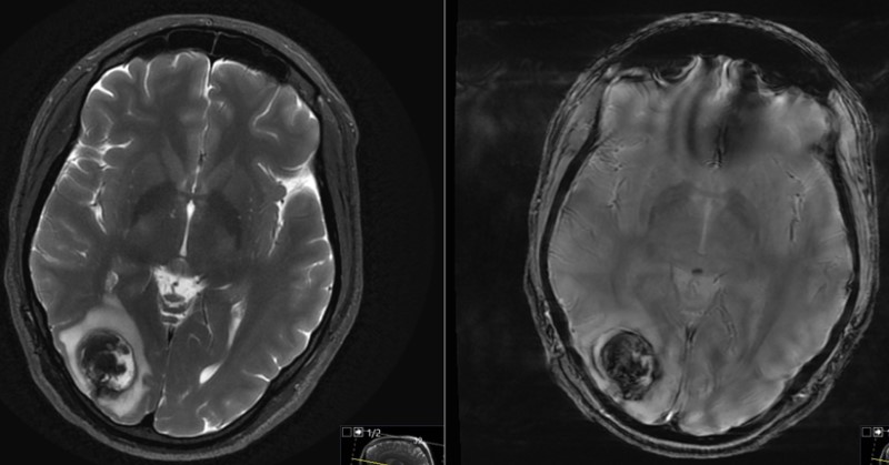
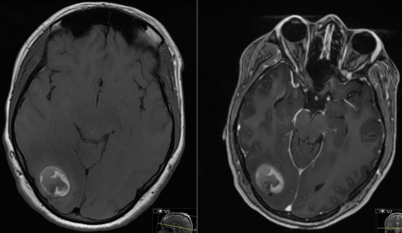
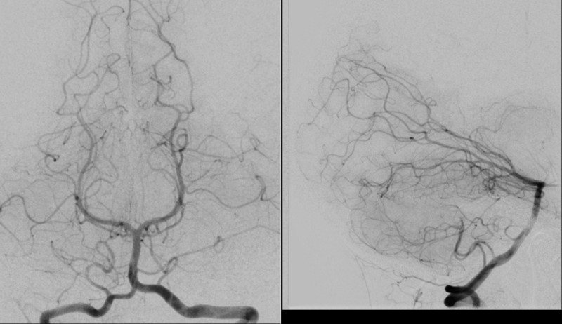
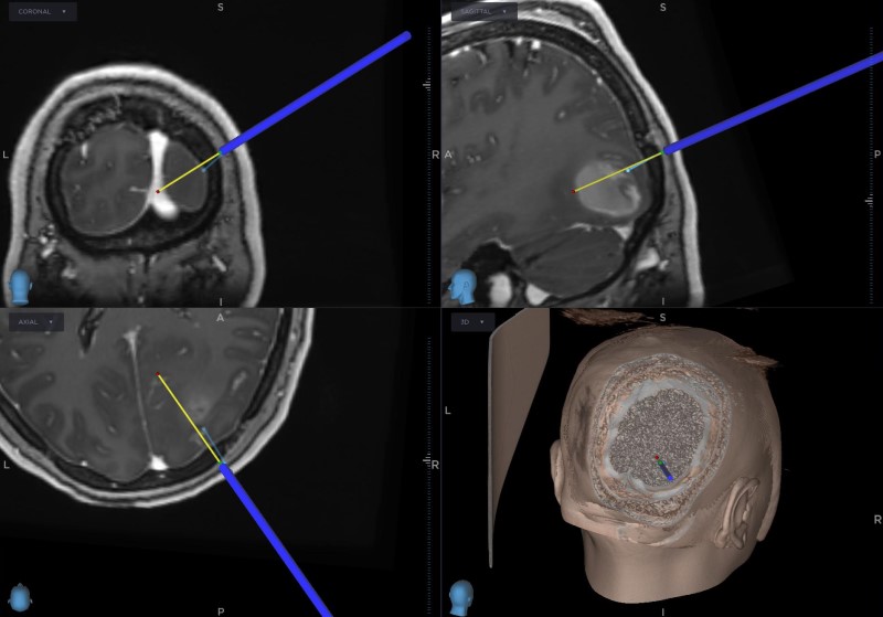
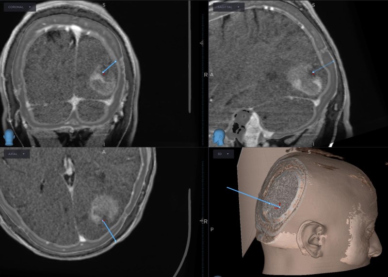
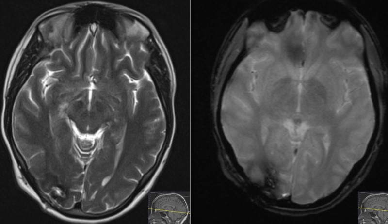
- Pre-Op
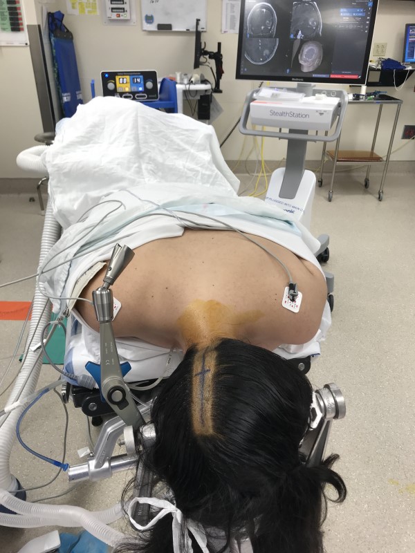
- Intra-op
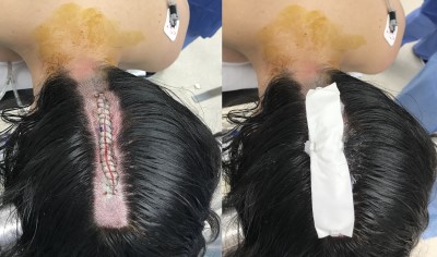
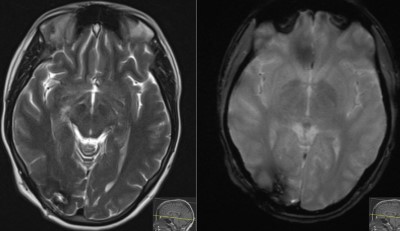
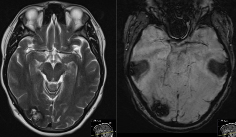
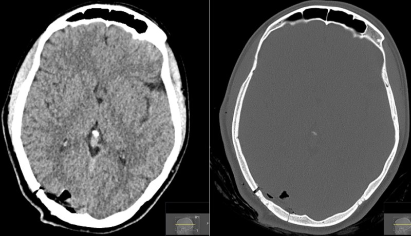
- Post-op


