This is a 61 year old male who undergoes an unrelated surgical procedure. Upon waking from surgery reports new pain, numbness, tingling in his forearms and hands and weakness of his legs.
No prior history of arm problems. He has no neck pain. Neuro exam with sensory loss in medial hands and radial forearm into axilla. He has proximal leg weakness. An urgent MRI c-spine was obtained.
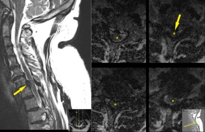
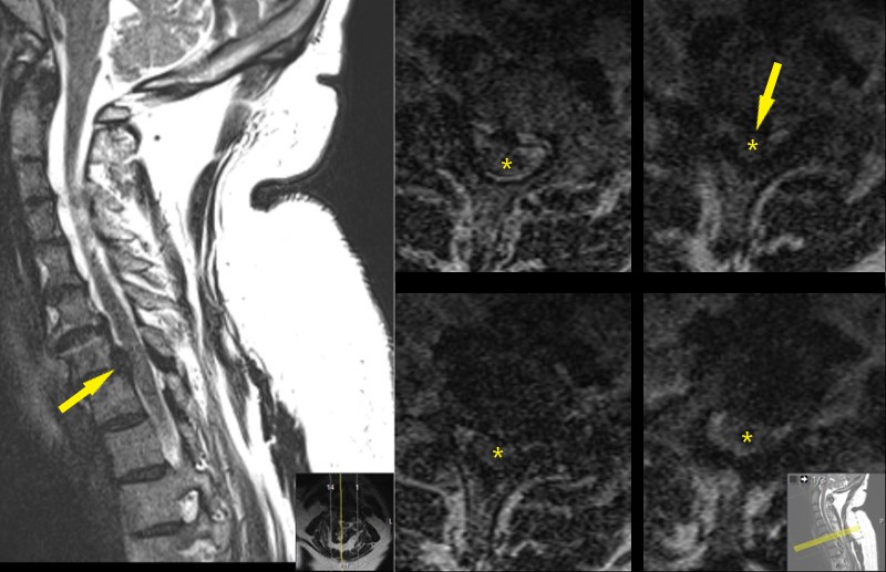
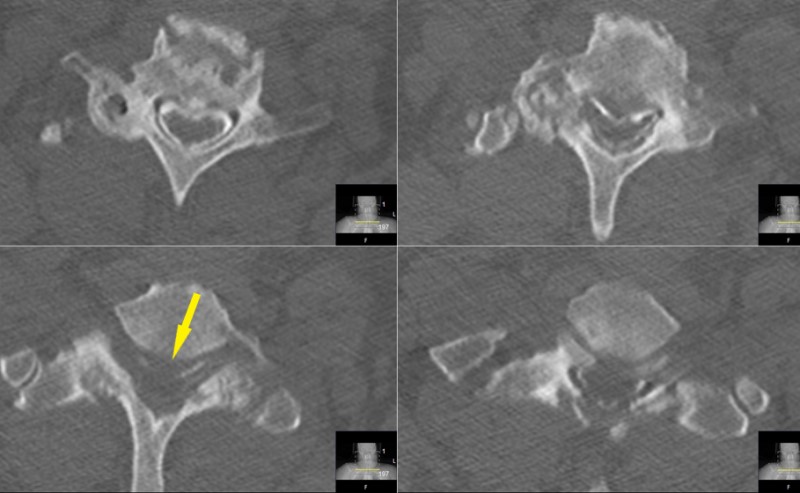
C-spine MRI. Midsagittal image (left) shows C7-T1 malalignment and disc displacement (yellow arrow). Axial images (left) are degraded by patient motion during the MRI scan. They show probable disc displacement and cord compression at C7-T1, but are inadequate to plan surgery. Disc displacement is shown by yellow arrow and spinal cord is designated by yellow star.
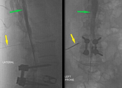
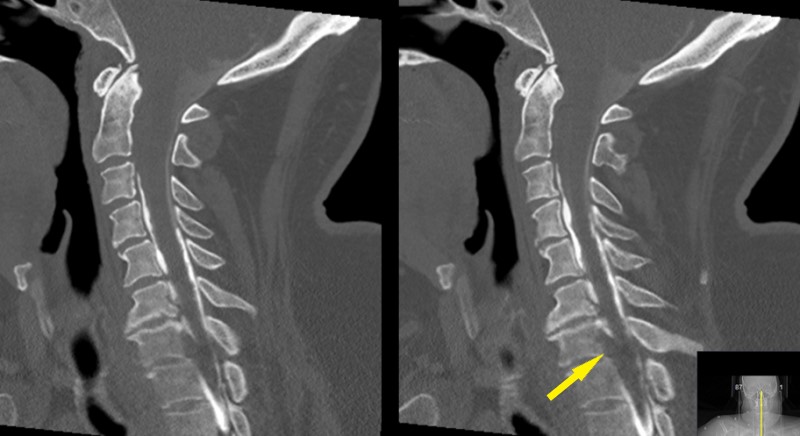
Myelogram - Fluoroscopic images of lumbar spine in the lateral (left) and AP (right) planes. Both images show results of recent lumbar spine surgery. Seen in both images is a needle that has been placed into spinal canal (lumbar puncture, yellow arrow). X-ray dye has been injected into the spinal canal and ascending up the back (green arrow). Note the dye outlining the lumbar nerve roots of the cauda equina.

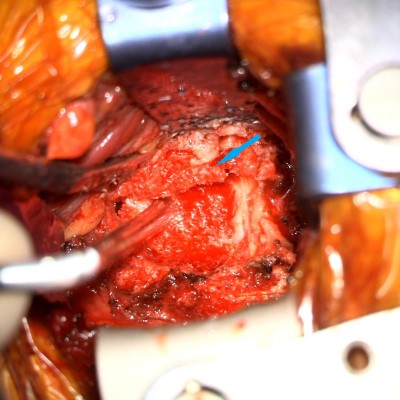
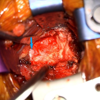
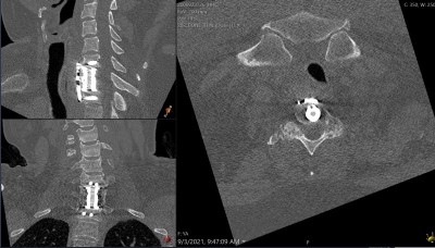
Intraoperative fluoroscopic images (lateral projections) taken during the C7 corpectomy and fusion procedure. Left image shows localizing needle in the C7-T1 disc space. Right images show the expandable interbody biomechanical device (aka cage) being applied. The upper image shows the top of the cage while the lower image shows the bottom.
- All
- Pre-Op
- Intra-op
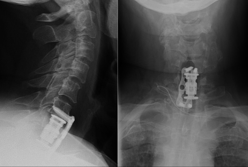
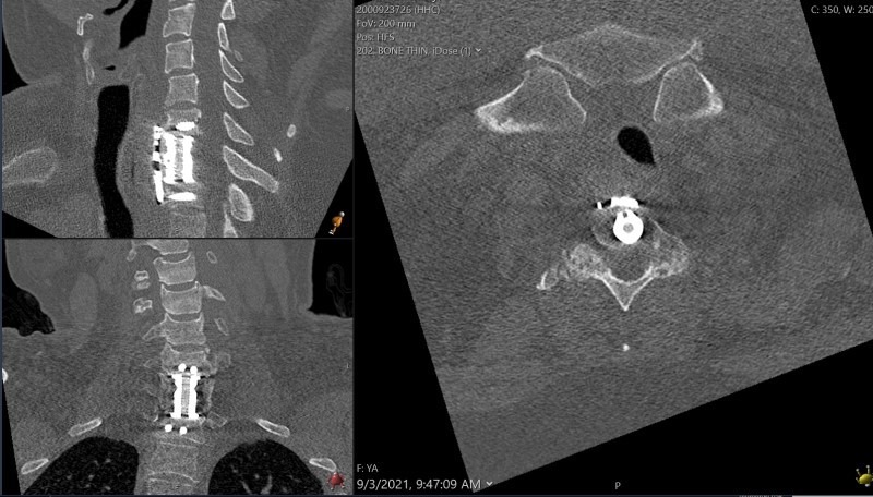
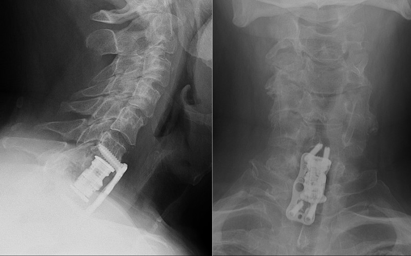
- Post-op


