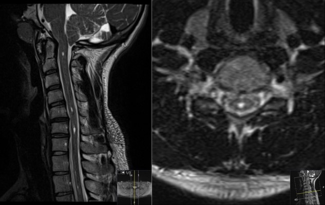
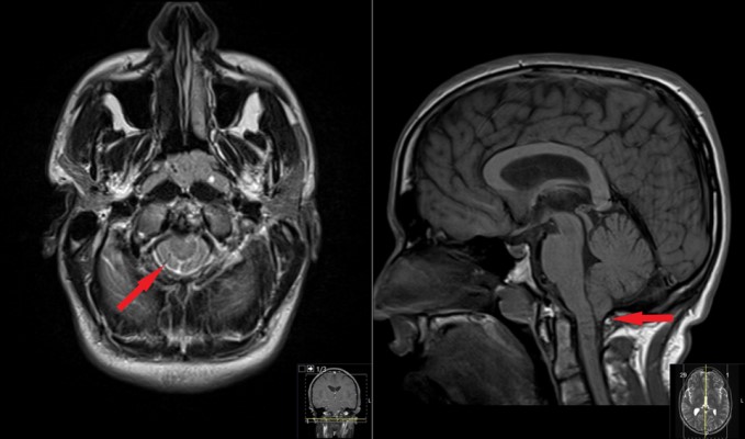
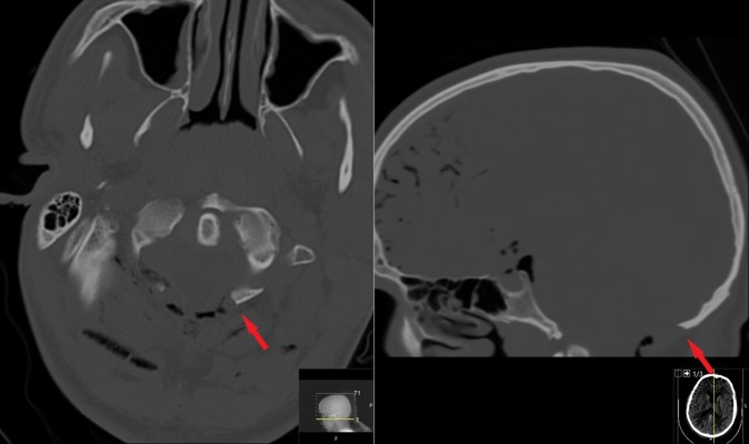
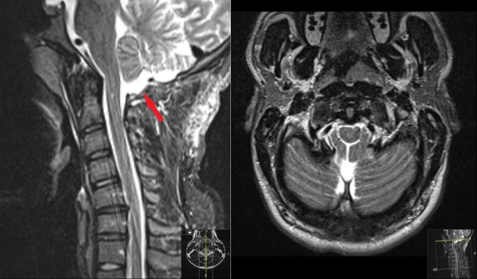
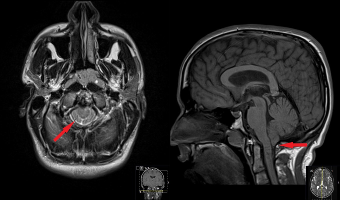
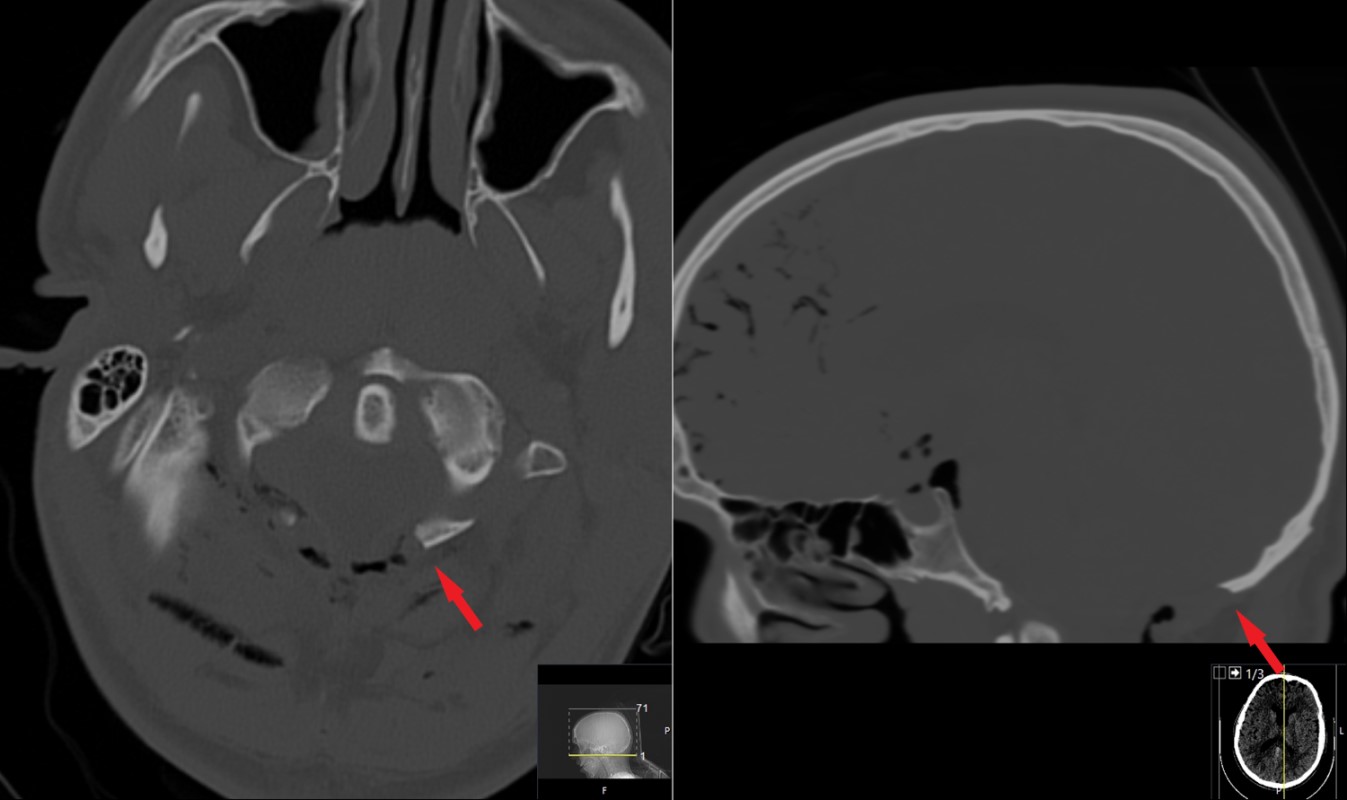
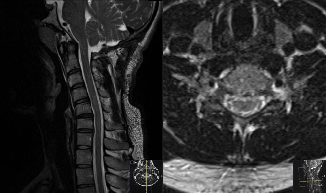
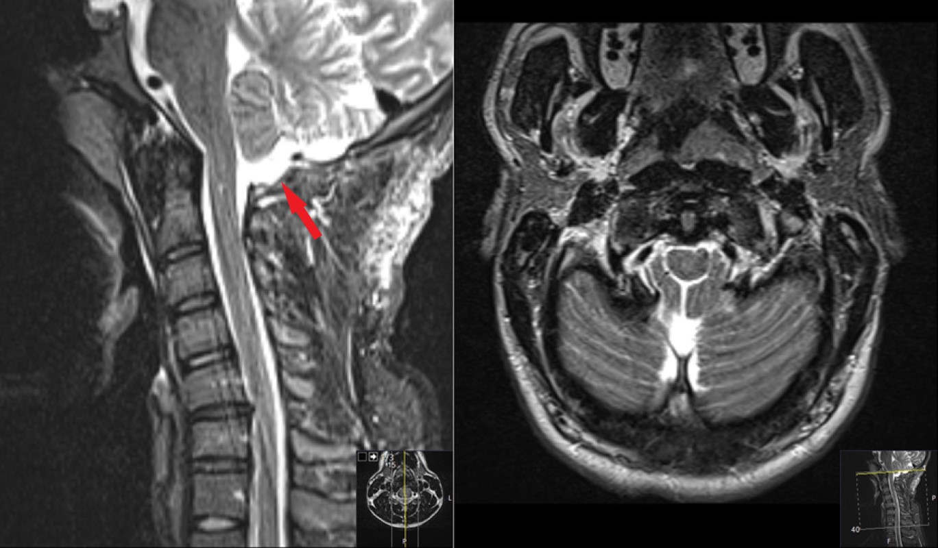
This is a 20 year old male who presented with headache with pressure in back of the head when lifting weights, coughing, and laughing for more than 1 year, and getting worse. His neurologic exam is unremarkable.
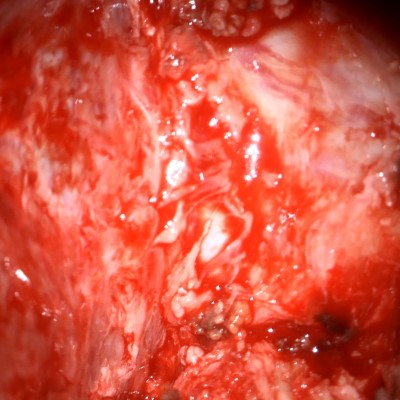
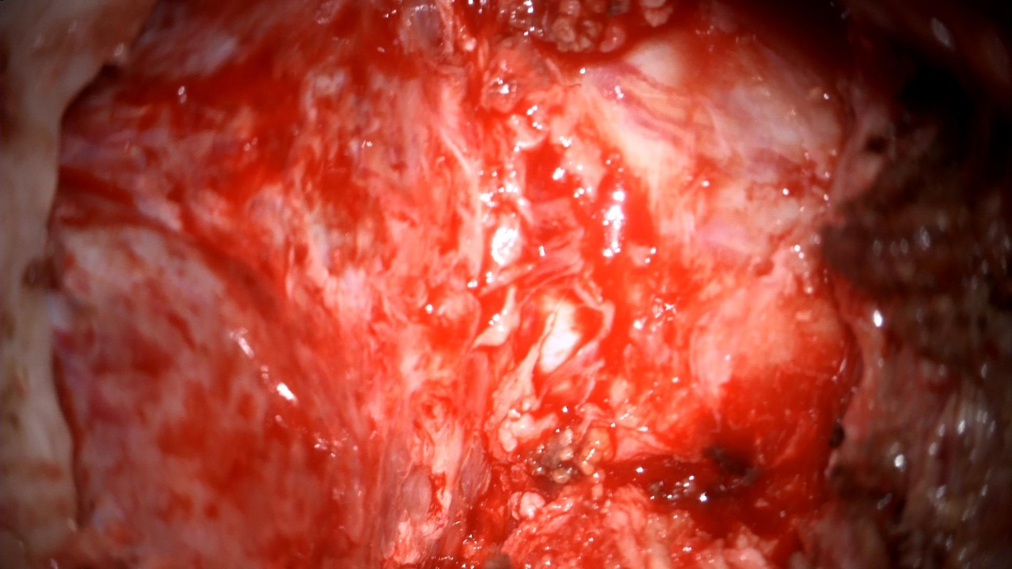
Intraoperative photo taken through the operative microscope. It shows the exposed dura of the posterior fossa and the upper cervical spine after removal of the suboccipital bone of the base of the skull and the lamina of C1. Orientation - this photo shows the back of the head and neck with the head being to the left of the image; right is to the top/left to the bottom.
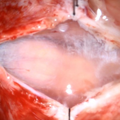
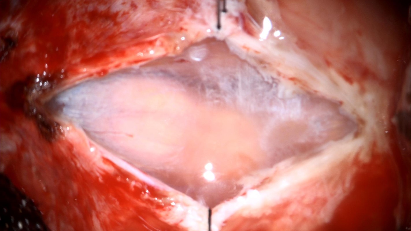
Intraoperative photo taken through the operative microscope. The dura has been opened, and the next membrane that covers the brain is exposed – the arachnoid mater. The cerebellum can be seen through the hazy membrane. Orientation - this photo shows the back of the head and neck with the head being to the left of the image; right is to the top/left to the bottom.
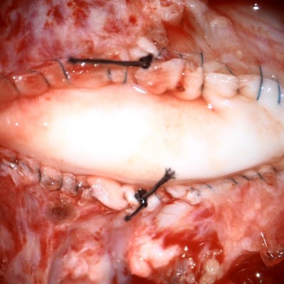
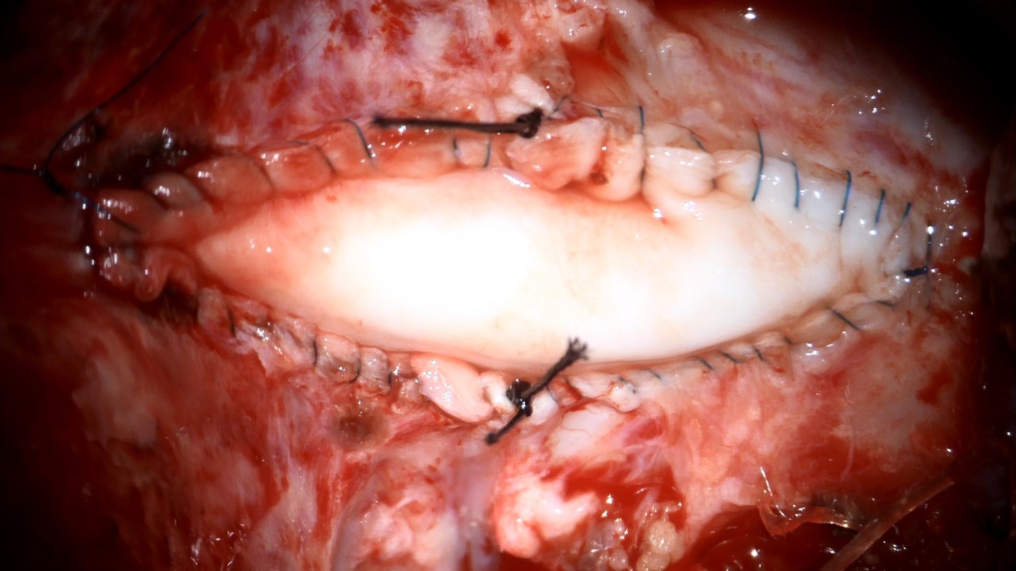
Intraoperative photo taken through the operative microscope. A patch has been sutured into the opening, covering the brain and preventing the escape of cerebrospinal fluid. Orientation - this photo shows the back of the head and neck with the head being to the left of the image; right is to the top/left to the bottom.
- All
- Pre-Op
- Intra-op
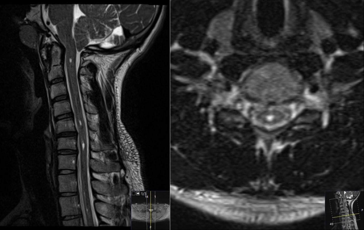
- Post-op
