Case: Craniotomy for a Glioblastoma multiforme (GBM)
This a case from a 64 year old male presenting with headache and visual changes. He had a headache for about 2 weeks getting stronger and was seeing flashing lights and losing sight in the left side of his vision.
- All
- Pre-Op
- Intra-op
- Post-op
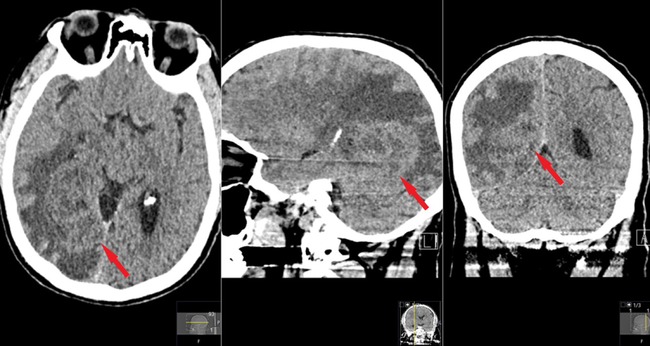
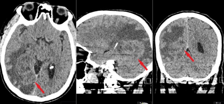 CT scan of the head showing an abnormality in the right parieto-occipital region of the brain (red arrows); demonstarted on axial (left), sagittal (middle) and coronal (right) planes.
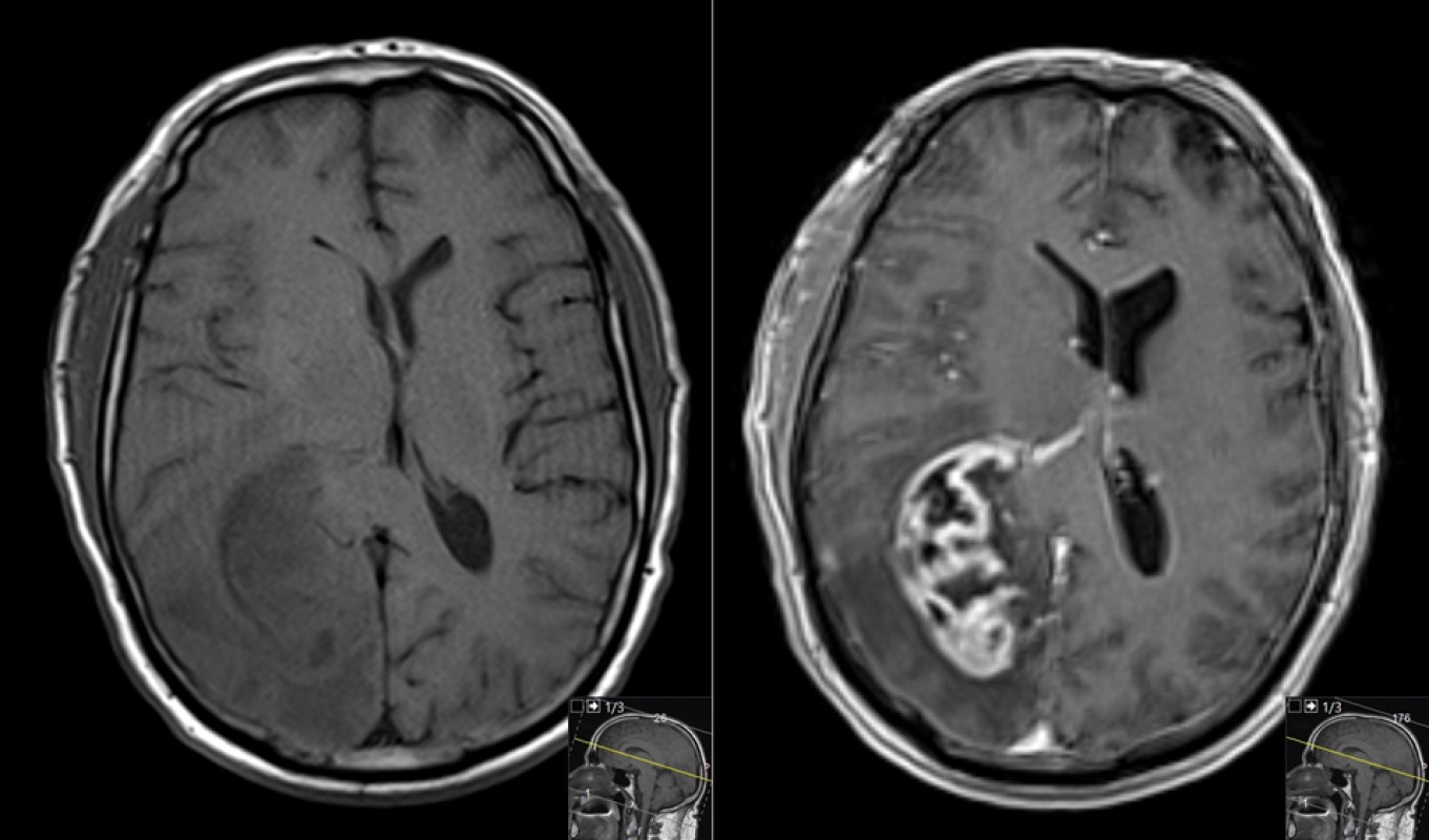
 Axial MR images performed without (left) and then with (right) intravenous contrast. The tumor, which is difficult to see on the pre-contrast images, becomes very obvious after the administration of contrast.
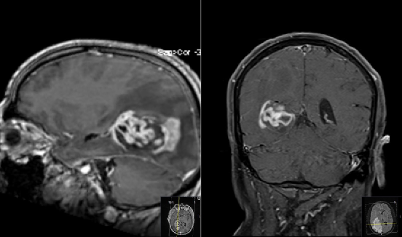
 Post-contrast MR images obtained in the sagittal (left) and coronal (right) planes further demonstrating the location and size of the tumor.
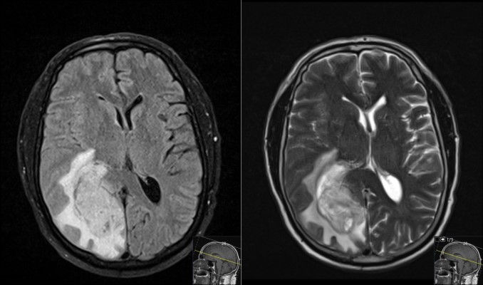 Axial FLAIR (left) and T2-weighted (right) MR images showing more characteristics of the tumor. Note the swelling around the tumor (aka edema, which is seen as a white intensity in the brain around the tumor) and the mass-effect the tumor is causing on the surrounding brain.
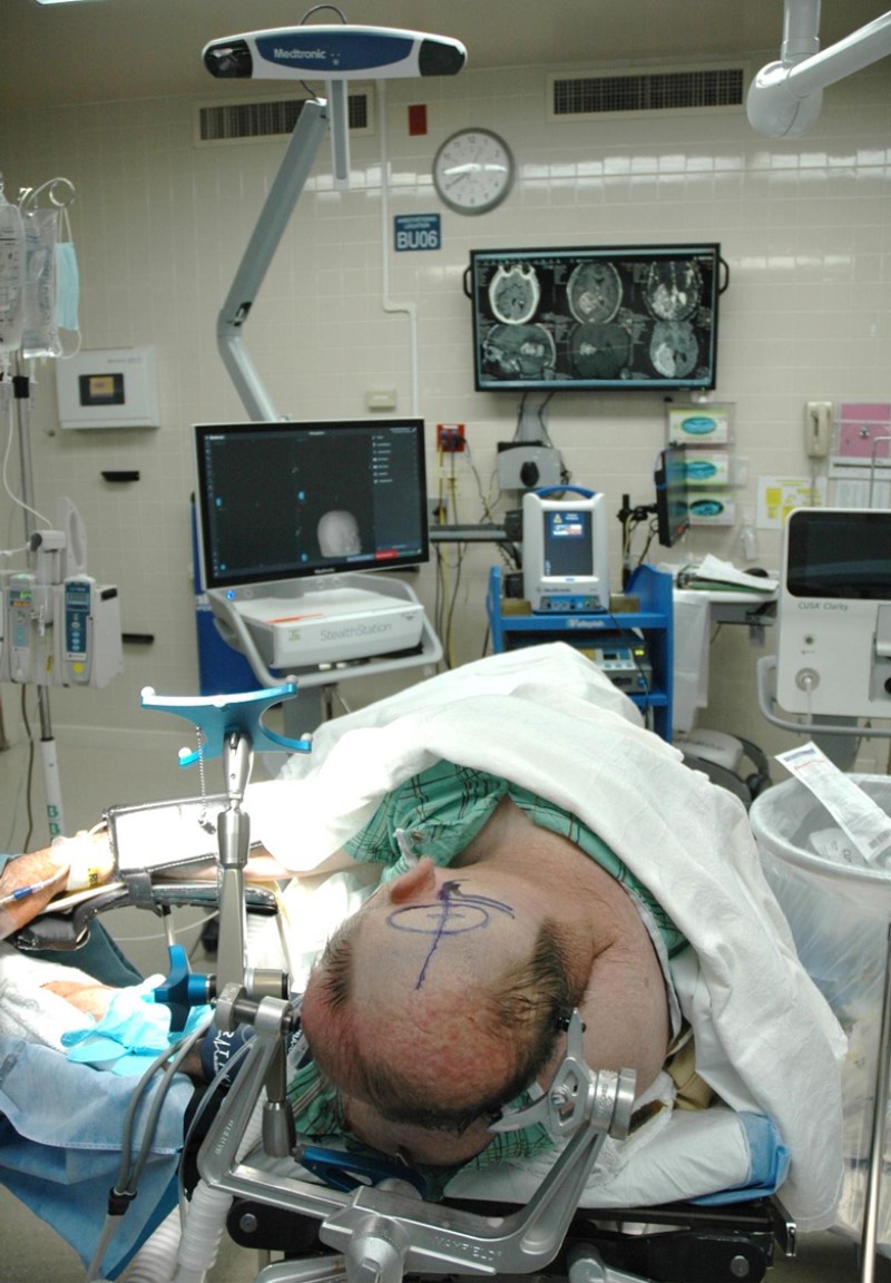
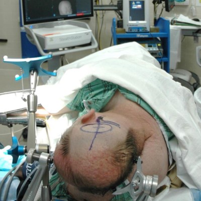 Intraoperative photo demonstrating patient positioning (in the left lateral position) and the planned skin incision; the outline of the tumor and other anatomy has been marked as well. Note the equipment in the foreground and background.
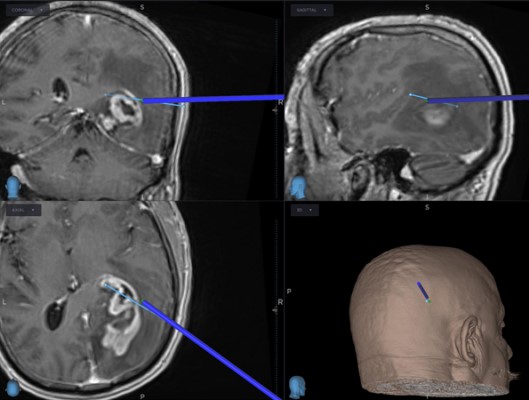 Image acquired from the navigation workstation demonstrating the planned approach to the tumor (thin, light blue line) and the virtual representation of an image-guided surgical instrument reaching the surface of the tumor (darker blue line).
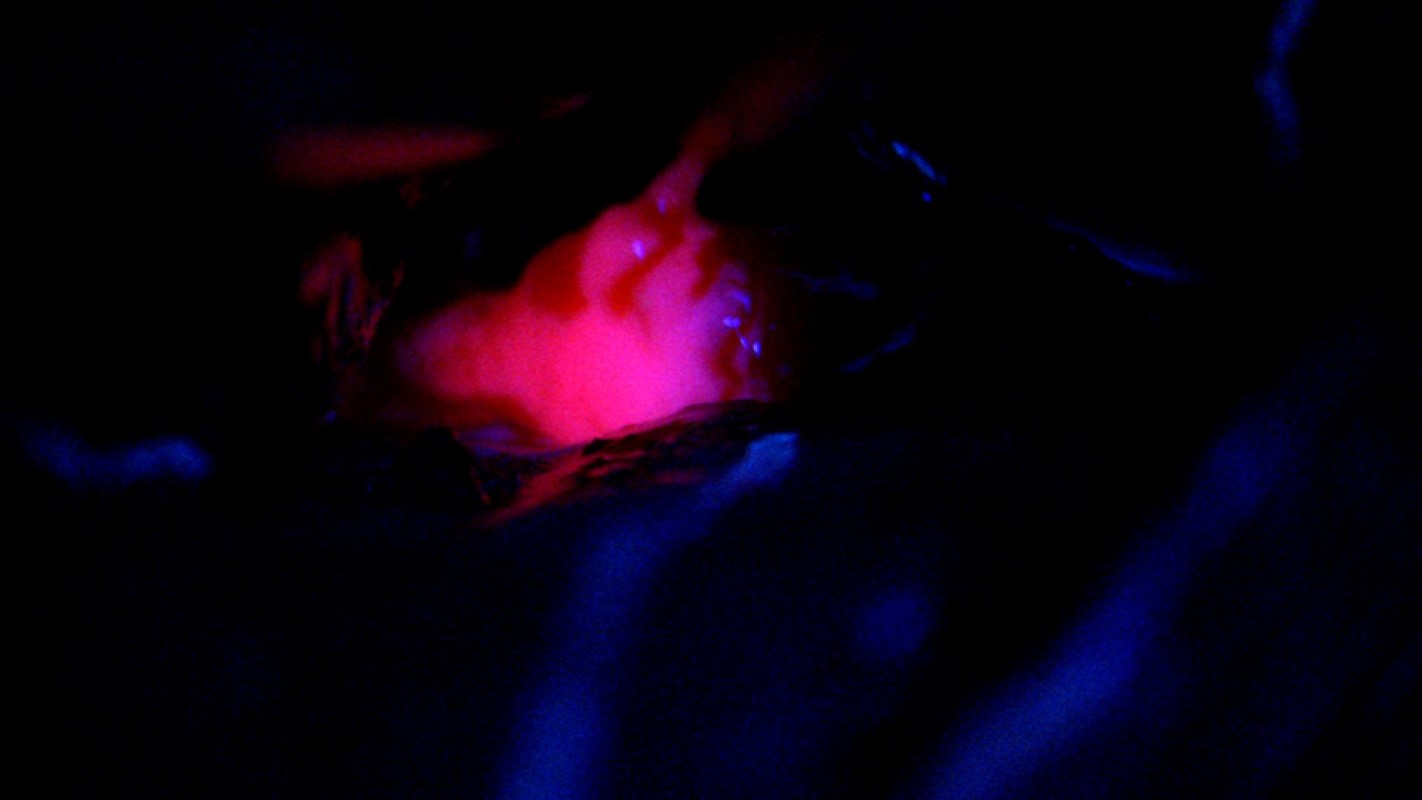
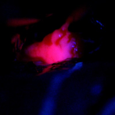 Fluorescence-guided tumor resection. Under blue light, the tumor glows pink demonstrating that access to the tumor has been achieved.
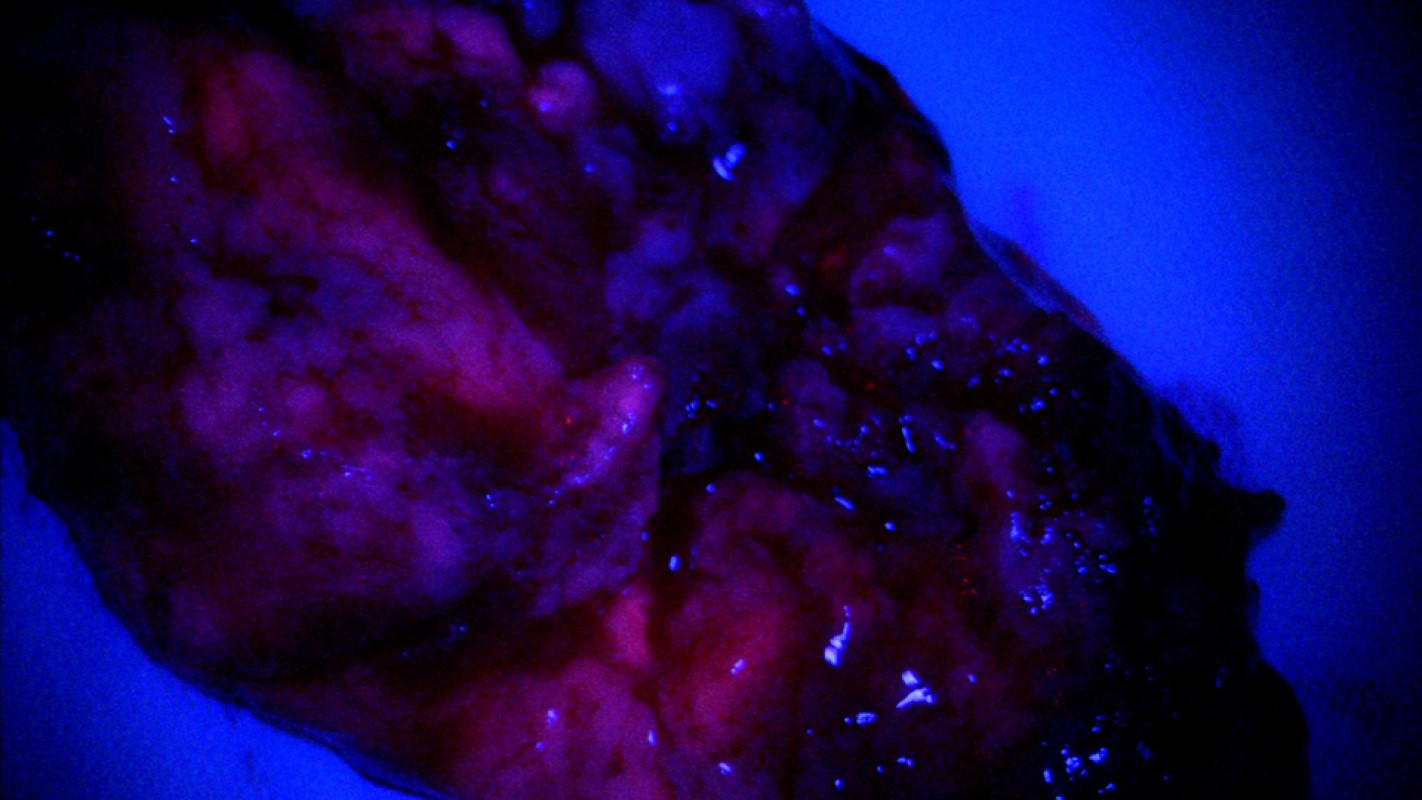
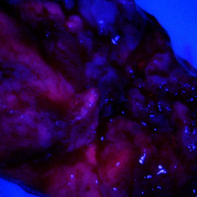 A large fragment of the tumor that has been removed and is being inspected under blue light. Note the continued fluorescence of the tumor (glowing pink).
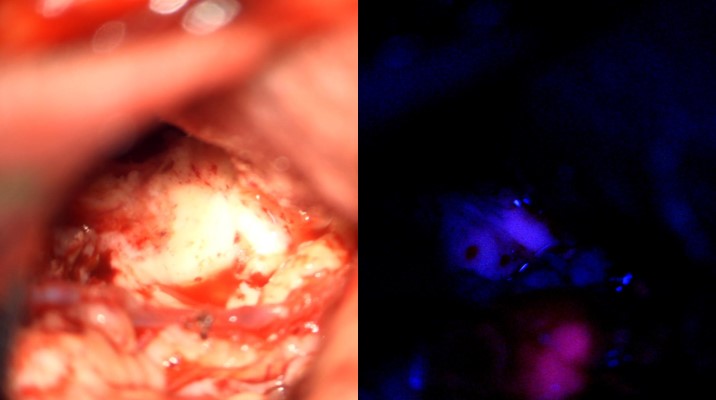 Intraoperative images acquired through the surgical microscope demonstrating fluorescence-guided tumor resection. The majority of the tumor has been removed, and under white light (left) there is no obvious residual tumor. When the light source is changed to blue light (right), a small fragment of tumor glows pink. This demonstrates complete resection in the upper portion of the surgical field, and a small residual in the lower field that can be removed.
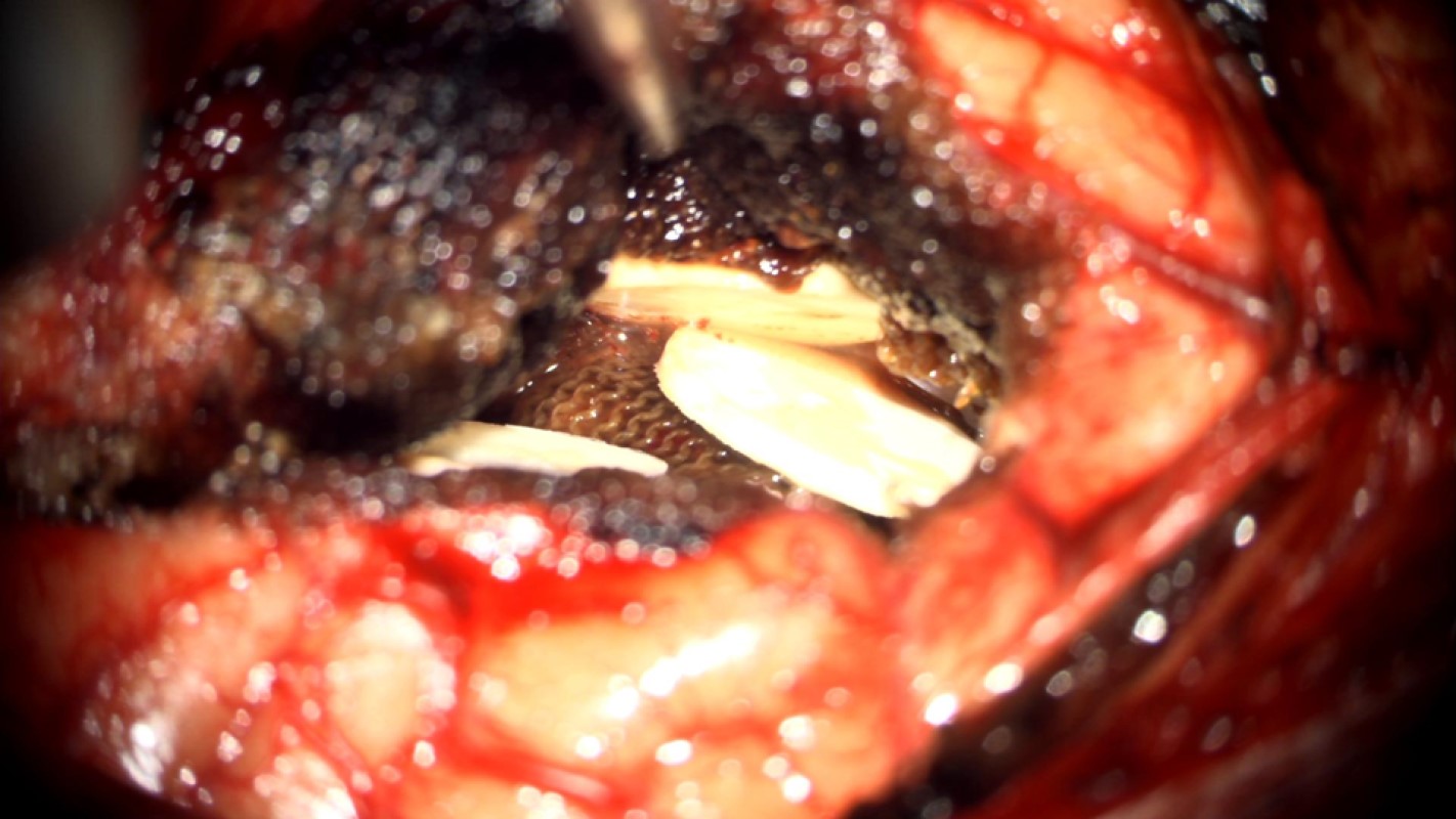
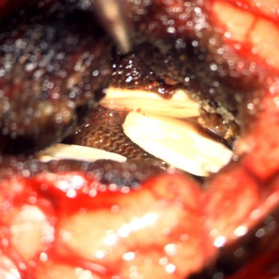 With the tumor completely resected, wafers that slowly release chemotherapy (carmustine, Gliadel) are placed into the resection cavity to begin treating microscopic disease.
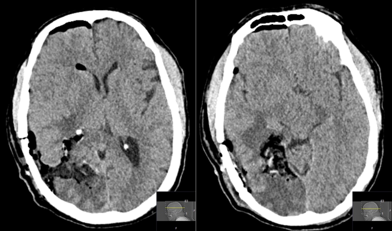
 Immediate postoperative CT of the head showing air and fluid in the resection cavity.
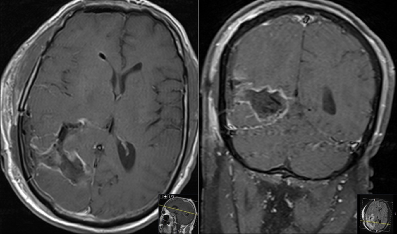
 MRI obtained 2 days after the surgery. Axial (left) and coronal (right) post-contrast images showing gross-total resection of the tumor.
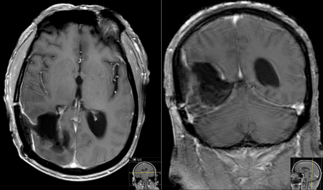
 MRI obtained 3 months after the surgery. Axial (left) and coronal (right) post-contrast images showing no residual or recurrence of the tumor.
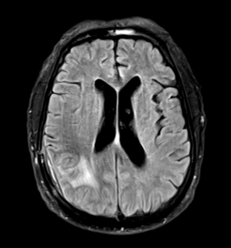
 Axial FLAIR MR image 3 months post-op. Compare with Figure 4 (left panel) showing marked reduction of edema in the region of the tumor.