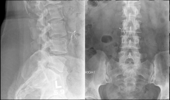
This a case of a 48 year old female who presented with back and right leg pain. The leg pain radiated to the bottom of the foot and was worsening despite conservative management. She also had numbness in her right foot as well as weakness in the right ankle.
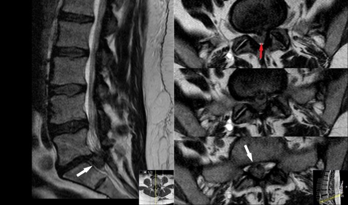
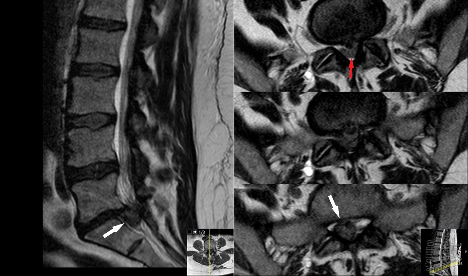
MRI lumbar spine. Sagittal image (left) shows disc herniation at L5/S1 level. Axial images (right) show disc herniation causing compression of the nerves within the spinal canal. The red arrow in the top image is pointing to the nerves within the spine. The bottom two images show the large disc herniation (white arrow) , notice that the nerves cannot be seen.
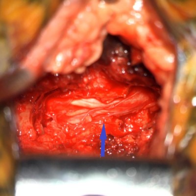
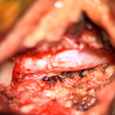
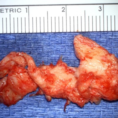
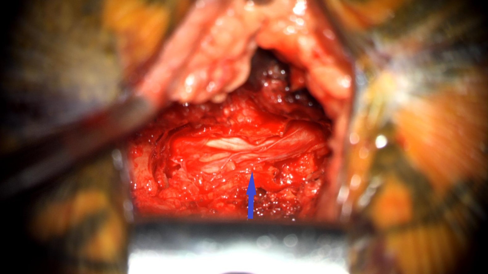
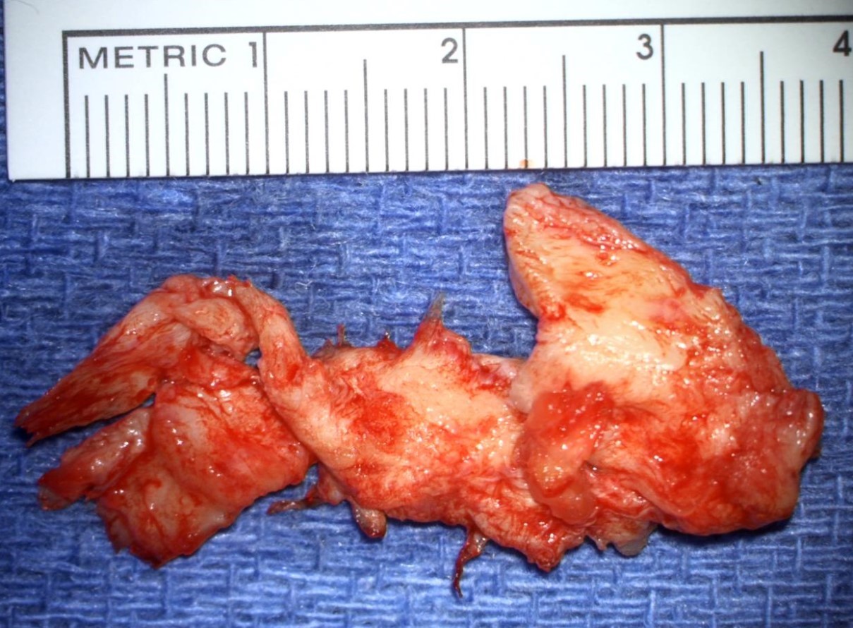
Surgical video showing removal of a large fragment of herniated disc material.
- All
- Pre-Op
- Intra-op
- Post-op
