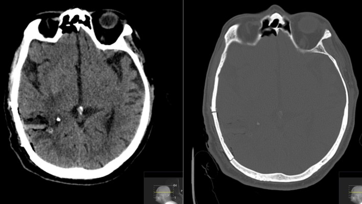
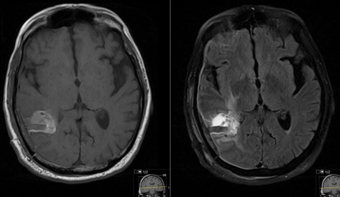
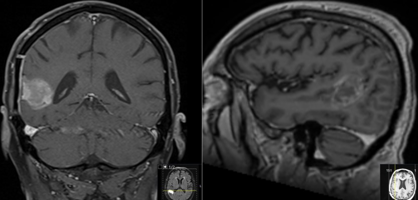
This is a 69 year old male with a known history of renal cell carcinoma who presented with cognitive decline and a neck mass.He had memory issues which were attributed to chemotherapy. He also was having new headaches. The neck mass was due to an infection. Neurologic examination was relatively unremarkable.
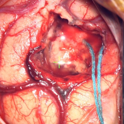
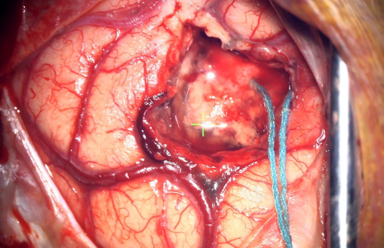
Intraoperative video obtained through the surgical microscope. The tumor has been debulked and dissected free from the surrounding brain. Video shows the final removal of the tumor.
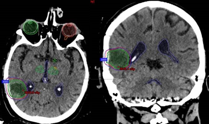
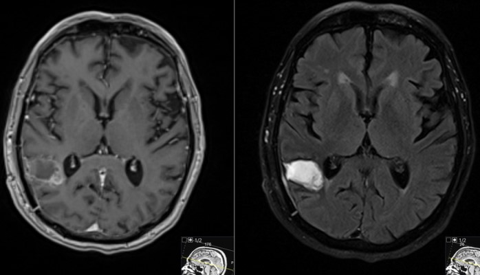
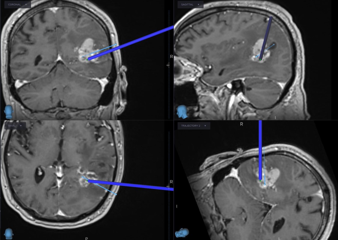
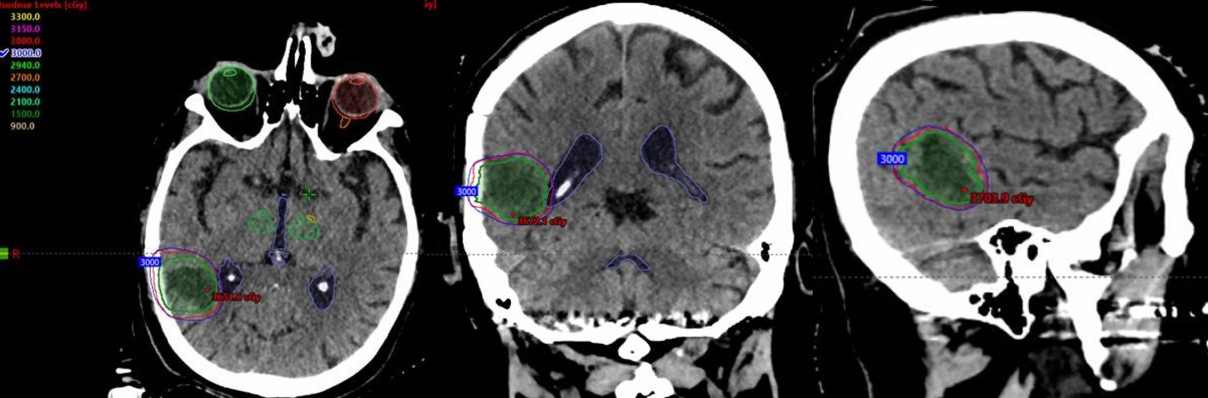
SRS planning images (merged CT & MRI) in the axial (left), coronal (middle) and, sagittal (right) planes with overlapping radiation dosing information. Note the conformity of the Stereotactic Radio Surgery (SRS) dose (demonstrated by the concentric rings around the tumor resection cavity) that treat the tumor bed without exposing the remainder of the brain to high doses of radiation.
- All
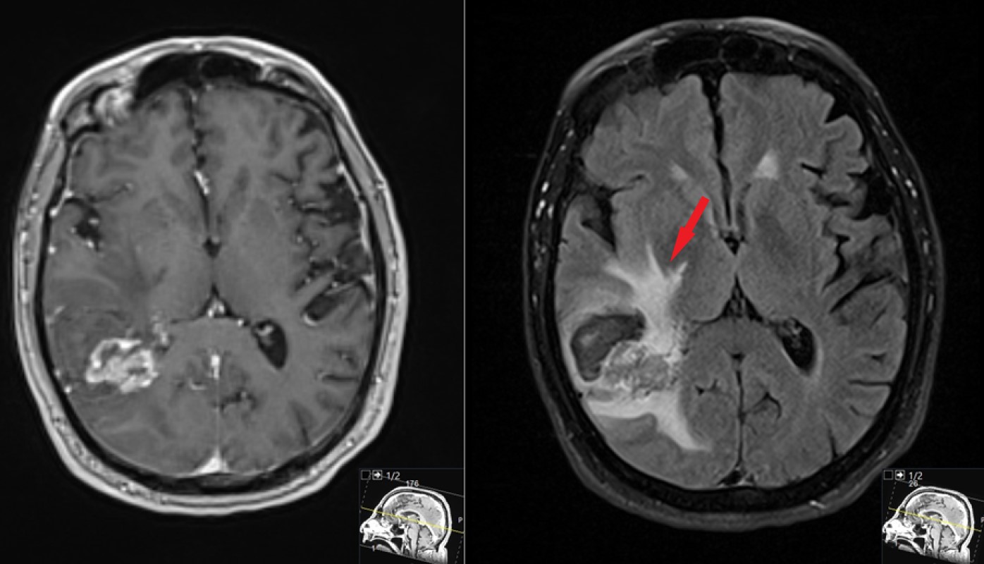
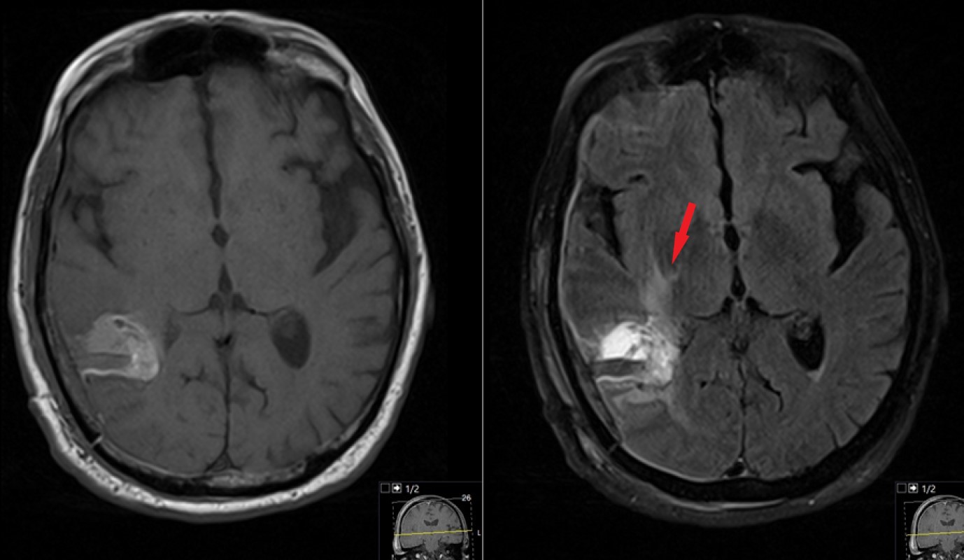
- Pre-Op
- Intra-op
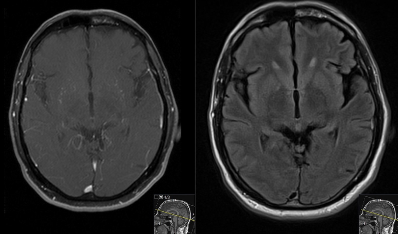
- Post-op
