This is a 28 year old female who presents with complaint of right-sided hearing loss and sense of fullness within her right ear. Her exam is consistent with conductive hearing loss in right ear.
  • All
  • Pre-Op
  • Intra-op
  • Post-op
Initial CT scan of head showing fluid within mastoid air cells and right middle ear (blue arrow).
Coronal reconstructions of temporal bone CT again showing fluid within the right mastoid air cells and middle ear. Also demonstrated is a defect (gap) in the floor of the cranium (blue arrow). Compare this with the left ear (right image) which shows air within the middle ear (black on CT).
Axial images from temporal bone CT also showing fluid within the right mastoid air cells and middle ear (blue arrow). Compare this with the left ear (right image) which shows air within the mastoid air cells and middle ear (black on CT).
Sagittal reconstructions of temporal bone CT again showing fluid within the right mastoid air cells and middle ear. Also demonstrated is a defect (gap) in the floor of the cranium (blue arrow).
Coronal MRI images (T2 on left, and T1 on right) showing brain tissue within the cranial floor defect (blue arrow).
Axial (left) and sagittal (right) MRI images ) also showing brain tissue within the cranial floor defect (blue arrow).
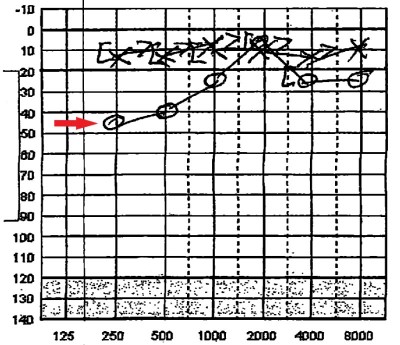
Audiogram demonstrating right-sided hearing loss. Normal, left-sided hearing is demonstrated by straight line of X’s across top of image. Right-sided hearing is represented by a series of O’s (red arrow). The drop of the line from the 10dB line is the amount of hearing lost; in this case 45dB at 250Hz and 25dB at 1000Hz & 4000Hz.
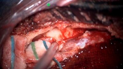
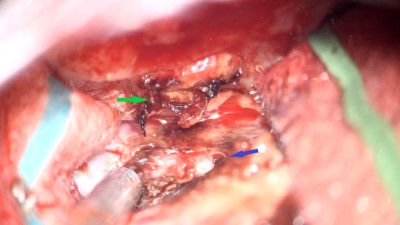
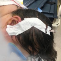
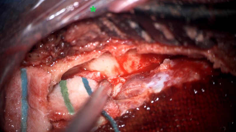
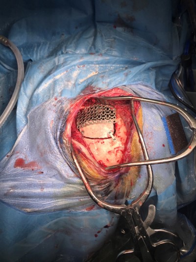
Intraoperative photo demonstrating the operative approach. A right temporal craniotomy has been made and the covering of the brain, the dura mater, is being dissected off of the base of the skull. Orientation: this is the right side of the head; the ear is just off the top of the frame (green star(*)), the nose is to the left, the back of the head the right, and the top of the head is toward the bottom.
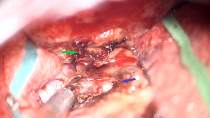
Intraoperative video demonstrating the dissection of the encephalocele. Note the band of tissue extending from the skull base defect on the top of the image through the dural defect toward the bottom. This is the encephalocele.
: Intraoperative photo demonstrating the pathology. The dura has been dissected free from the skull base and the encephalocele has been resected. The defect in the skull base is at the top of the field (green arrow) and the defect in the dura is toward the bottom (blue arrow).
Intraoperative photo of the skull base repair. The cranial bone flap has been used to cover the defect in the middle fossa floor. A small amount of bone cement (Hydroset) has been used to seal the defect and secure the bone in place.
A photo of the operative field at the completion of the skull repair. The remaining bone flap has been replaced along with a titanium mesh cranioplasty to cover the craniotomy that was made to access and repair the encephalocele and skull base defect.
A photo of the repair. The remaining bone flap has been replaced along with a titanium mesh cranioplasty to cover the craniotomy that was made to access and repair the encephalocele and skull base defect.
Photo taken at the completion of the procedure showing the location of the incision on the right side of the head and the dressing applied to a minimal-shave brain surgery.
Postoperative CT head showing the repair in the axial, coronal, and sagittal planes (left-to-right). Demonstrated are the titanium mesh cranioplasty (blue arrows) and the patient’s own cranial bone used to repair the skull base defect (yellow arrows).