This is a 63 year-old woman presented with headache. She has no visual change or complaint. She reports prior history of a pituitary tumor.
Her examination unremarkable. Visual field full to confrontation. Her labs are unremarkable. Her Prolactin level was 9.0 ng/mL (normal 2 – 20).
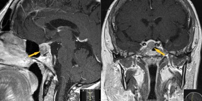
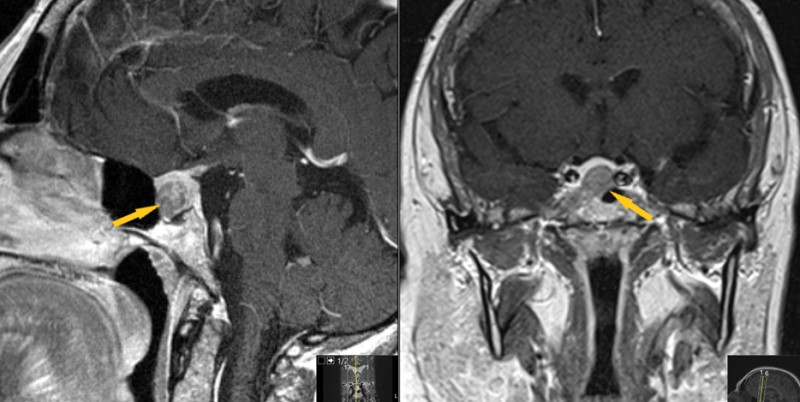
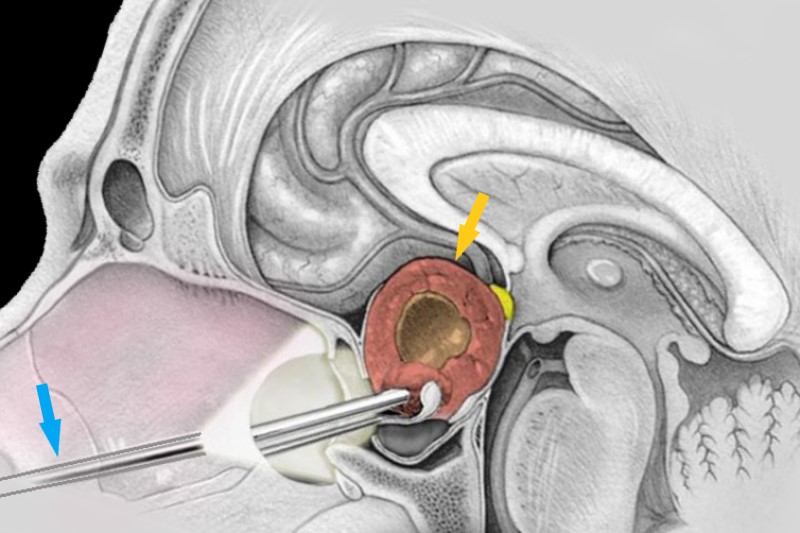
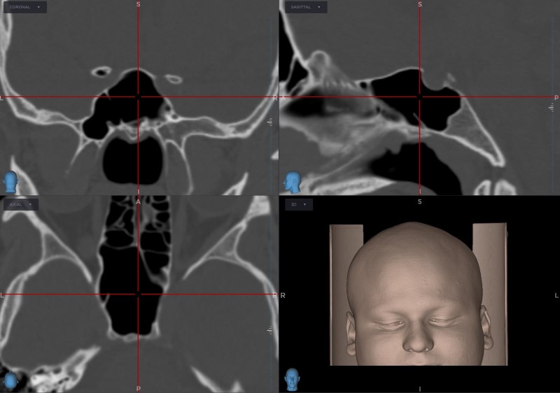
Recent MRI brain with pituitary protocol: Postcontrast T1 images in sagittal (left and axial (right) planes. Shows pituitary lesion within the sella turcica elevating and compressing the pituitary gland. Tumor is round, grey lesion (gold arrow) and pituitary gland is white curved line on top of lesion.
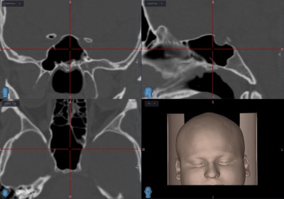
Intraoperative video captured form the navigation system. It shows how instruments are tracked in real time by the navigation workstation and how the information is relayed to the surgeon accessing the sphenoid sinus.
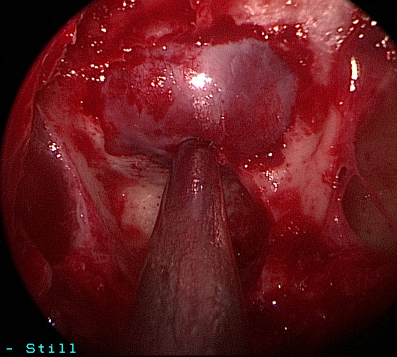
Intraoperative video showing resection of the tumor with a pituitary ring curette. The tumor is soft is carefully removed piece by piece.
Intraoperative video showing resection of the tumor with a pituitary ring curette. The tumor is soft is carefully removed piece by piece.
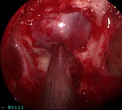
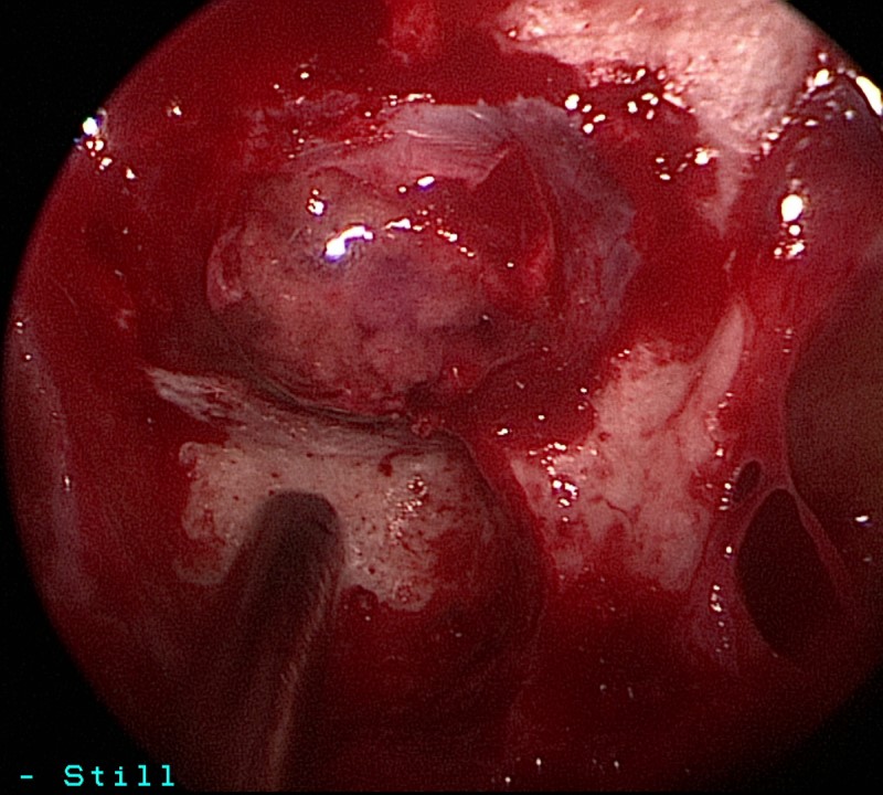
Intraoperative video taken after the tumor has been largely resected. The endoscope is advanced into the sella turcica and the right wall of the sella is inspected. The glistening pink object is the right internal carotid artery. The tan-ish tissue is pituitary gland. There is still a small piece of tumor between the carotid and the gland that needs to be removed. Also visible is the pink supra-sellar arachnoid membrane hanging down into the sella. This is the great advantage of the endoscope- the ability to look within the resection cavity.
Her examination unremarkable. Visual field full to confrontation. Her labs are unremarkable. Her Prolactin level was 9.0 ng/mL (normal 2 – 20).
- All
- Pre-Op
- Intra-op
- Post-op


