This is a 65 year-old male who presented to the ER after multiple falls. He describes progressive hand weakness and loss of dexterity along with difficulty walking resulting in multiple falls recently.
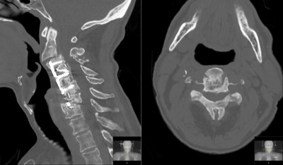
He has a history of prior anterior cervical spine surgery approximately 13 years prior. The neurological examination shows significant weakness of all extremities, more in hands and proximal legs. Hyperreflexia with abnormal reflexes. Diminished fine motor movement in hands. He gets a CT and MRI in ER.
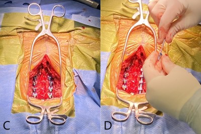
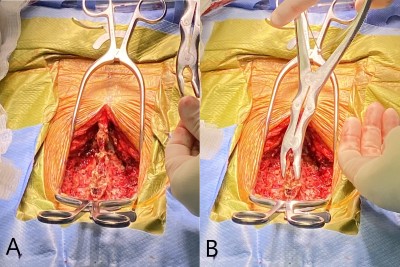
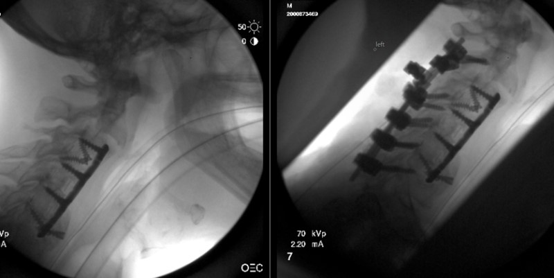
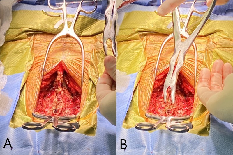
Images obtained during surgery. Patient is positioned face down (prone). Head is at bottom of image. (C) Shows the posterior cervical spine after completion of the laminectomy and with lateral mass screws in place. (D) Shows rod in place on patient’s left side and right-side rod being assembled in the surgeon’s hands.
- All
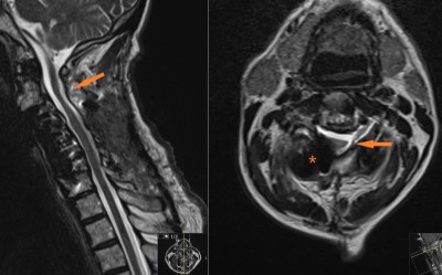
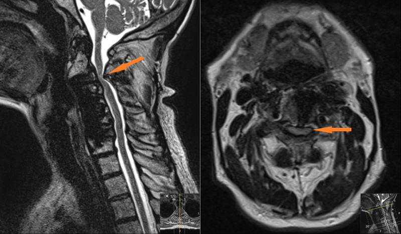
- Pre-Op
- Intra-op
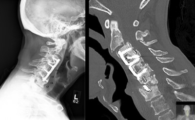
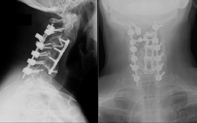
- Post-op

