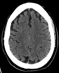
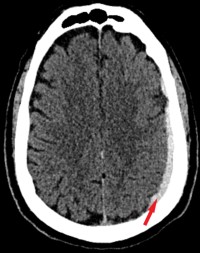
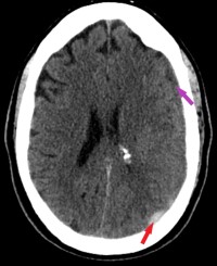
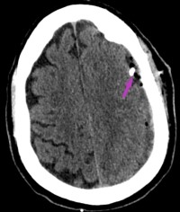
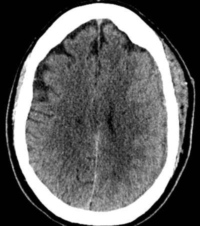
This is a 70 year old male presents to ER after seizure. He has no history of seizure. The seizure occurred at dinner table. He had generalized shaking with loss of consciousness, witnessed by wife. The patient is without memory of the event. He complains of headache. He has no history of trauma. The patient has history of cardiac stents placed 3 years prior and takes aspirin, 81mg daily, and clopidogrel (Plavix) 75 mg daily.
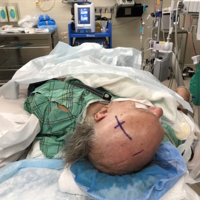
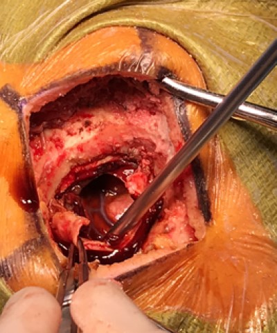
Video taken in the OR. The craniotomy has been performed and the dura has been opened. An electro-cautery is used to perforate the exposed subdural membrane. The chronic subdural hematoma then is spontaneously expressed under some pressure. The underlying brain is then seen.
- All
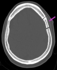
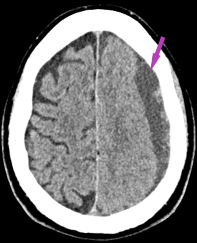
- Pre-Op
- Intra-op
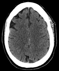
- Post-op


