Image Guided Surgery
Image-guided surgery is a term describing any surgical procedure in which the surgical team uses tracked surgical instruments in conjunction with preoperative or intraoperative images in order to directly or indirectly guide the procedure. Image guided surgery systems typically use infrared or visible light cameras, or electromagnetic fields to capture and relay the patient's anatomy and the surgeon's precise movements in relation to the patient. That information is then viewed on computer monitors in the operating room in real-time as a virtual representation of the surgical instrument in conjunction with the imaging study. One way to describe it more simply is the analogy of GPS for an operation. Rather than seeing a virtual car on a map and a depiction of the rout to take, the surgical instruments can be seen overlying an MRI or CT scan, and a surgical plan can be developed and followed.
Image-guided surgery was originally developed for brain surgery to help neurosurgeons perform safer and less invasive procedures. It allows surgeries to be performed more safely by guiding the surgeon to the surgical target (like a tumor) while avoiding normal tissues. It also has the advantage of performing surgery more quickly with smaller incisions allowing better outcomes and a more rapid recovery. While image guidance is not needed for all surgeries, it has become a recognized standard of care in many operative procedures in cranial neurosurgery, and more recently, due to advances in intra-operative imaging technology, has been used in spinal applications as well.
Dr. Hornyak and his surgical team use the most advanced Neurosurgical navigation system available, the Stealth S8, from Medtronic. It is used for most brain tumor surgeries, including brain biopsy procedures. When it is used in conjunction with the O-arm, an intraoperative 3D imaging device, image-guidance can be used in spine surgery as well.

Figure 1: This image shows a representation of a car (the blue arrow) on a map and includes a route to follow to a predetermined destination.
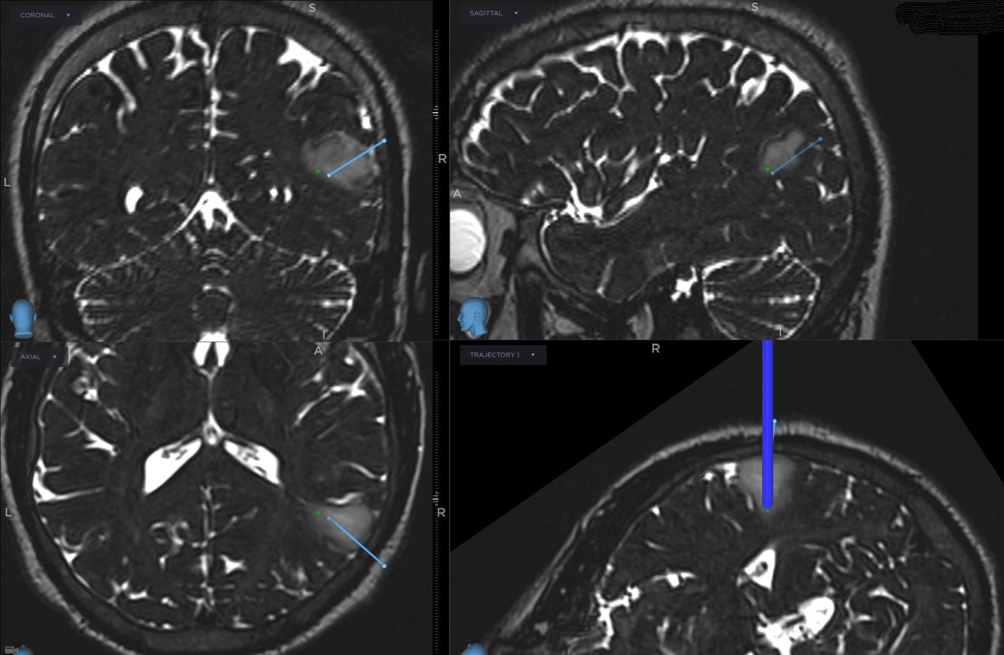
Figure 2: This image shows a representation of a surgical instrument (the thick blue line in bottom right pane) on a pre-operative MRI of a patient with a brain tumor (the whitish area) and includes a predetermined trajectory to most safely access the lesion (the thin blue line).